Review
. 2015 Jan;235(2):266-76.
doi: 10.1002/path.4462.

Respiratory syncytial virus (RSV) and its propensity for causing bronchiolitis

Affiliations
Review

Respiratory syncytial virus (RSV) and its propensity for causing bronchiolitis

Raymond J Pickles et al. J Pathol. 2015 Jan.

Abstract

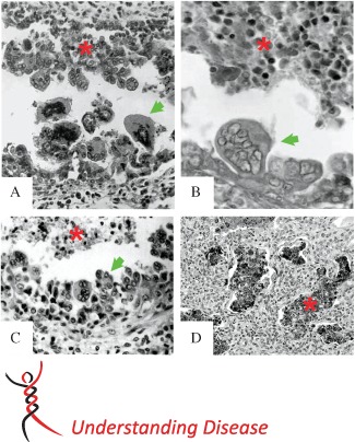
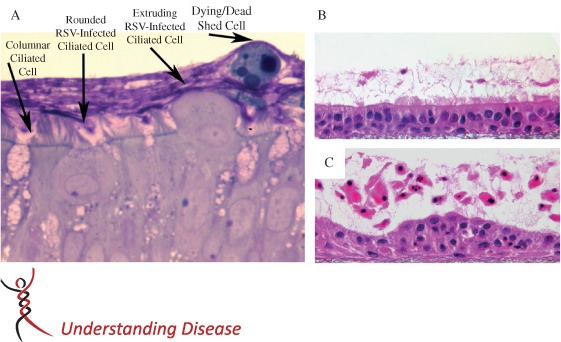
Infants and young children with acute onset of wheezing and reduced respiratory airflows are often diagnosed with obstruction and inflammation of the small bronchiolar airways, ie bronchiolitis. The most common aetological agents causing bronchiolitis in young children are the respiratory viruses, and of the commonly encountered respiratory viruses, respiratory syncytial virus (RSV) has a propensity for causing bronchiolitis. Indeed, RSV bronchiolitis remains the major reason why previously healthy infants are admitted to hospital. Why RSV infection is such a predominant cause of bronchiolitis is the subject of this review. By reviewing the available histopathology of RSV bronchiolitis, both in humans and relevant animal models, we identify hallmark features of RSV infection of the distal airways and focus attention on the consequences of columnar cell cytopathology occurring in the bronchioles, which directly impacts the development of bronchiolar obstruction, inflammation and disease.

Keywords: acute lung disease; airway obstruction; bronchiolitis; respiratory syncytial virus.


Figures

Figure 1
Figure 1
Histopathology of RSV‐infected small airways. Previously published histology images of common small airway lesions identified post mortem in human infant lungs infected with RSV. All examples describe disruption of the distal airway epithelium, with giant cell or polypoid formation (green arrows) and sloughed or protruding RSV antigen‐positive epithelial cells retained in the bronchiolar lumen (red asterisk). (A) A 1959 RSV case, showing bizarre dearrangement of the small airway epithelium, with multi‐nucleated, polypoid epithelial cells casting off into the airway lumen. Figure reproduced from 25. ‘Immunofluorescent evidence of respiratory syncytial virus infection in cases of giant‐cell bronchiolitis in children’, Vol. 89, Pages 343–347. Copyright © 1965 The Pathological Society of Great Britain and Ireland. This material is reproduced with permission of John Wiley & Sons, Inc. (B) A 1949 RSV case, demonstrating papilliary projections and intrabronchiolar syncytia contributing to the intraluminal cellular debris. Figure reproduced from 27. Reprinted by permission from Macmillan Publishers Ltd: Modern Pathology, ‘The histopathology of fatal untreated human respiratory syncytial virus infection’, Copyright 2007. (C) A pre‐1988 RSV case, showing virus‐induced airway epithelium injury, characterized by uneven proliferation of epithelial cells with protrusions entering into the bronchiolar lumen, creating a polypoid appearance. Figure reproduced from 30: KA Neilson and EJ Yunis, Fetal & Pediatric Pathology, [1990; 10 (4): 491–502], Copyright © 1990, Informa Healthcare. Reproduced with permission of Informa Healthcare. (D) Immunohistochemical detection of RSV antigen in exfoliated bronchiolar epithelial cells clogging the lumens of small airways. Reproduced from 28. Welliver et al, ‘Respiratory syncytial virus and influenza virus infections: observations from tissues of fatal infant cases’, The Pediatric Infectious Disease Journal, Vol. 27, Suppl. 10, pages S92–S96, with permission. Original magnifications = (A) × 150; (B) × 250; (C) × 650; (D) × 40
Figure 2
Figure 2
RSV infection of differentiated cultures of human airway epithelium. (A) Histological cross‐section of HAE cultures, with ciliated columnar cells transitioning from columnar to rounded cell morphology during RSV infection and followed by extrusion of the infected cells from the epithelium and into lumenal surface secretions. After shedding, detached epithelial cells rapidly become apoptotic, while being transported across the culture surface by beating cilia located on underlying and non‐infected ciliated cells. Cultures were fixed in perfluorocarbon impregnated with osmium tetroxide and embedded in plastic. Section shown was counterstained with Richardson's. (B, C) Histological cross‐sections of HAE, 3 days after inoculation of UV‐inactivated RSV (B) or RSV (C), demonstrating how RSV infection of ciliated cells results in disruption of the epithelium, atypical epithelial cell morphology and robust shedding of epithelial cells into lumenal surface secretions. Detached epithelial cells showed morphological evidence of pyknosis, karyorrhexis and karyolysis, indicative of an apoptosis‐like cell death. Cultures were fixed in Omnifix to preserve lumenal secretions, embedded in paraffin and the sections counterstained with haematoxylin and eosin (H&E) 71. Republished with permission of the American Society for Clinical Investigation, from ‘RSV‐encoded NS2 promotes epithelial cell shedding and distal airway obstruction’, The Journal of Clinical Investigation, RM Liesman et al, Vol. 124, Iss. 5, Copyright 2014; permission conveyed through Copyright Clearance Center Inc.
Figure 3
Figure 3
Early epithelial cell cytopathology and inflammation in hamster bronchioles infected by PIV3 or PIV3–NS2, compared to neonatal lamb bronchioles infected by RSV. Histological cross‐sections of bronchiolar airways from hamsters (A, B) infected by PIV3 (A) or PIV3–NS2 (B), and bronchiolar airways from neonatal lambs infected by RSV. Reproduced from 42, ‘Perinatal Lamb Model of Respiratory Syncytial Virus (RSV) Infection’, by Derscheid and Ackermann, 2012, licensed under CC‐BY 3.0. (C). PIV infection results in modest epithelium cytopathology, with robust neutrophil‐rich inflammatory cell infiltration into peribronchiolar (green arrow) and intralumenal (white arrow) compartments, resulting in moderate loss of airway patency. In contrast, hamster and lamb bronchioles infected by PIV3–NS2 or RSV, respectively, show strikingly similar consequences of infection: disruption and dearrangement of the epithelium, with epithelial cells protruding and shedding into the airway lumen (yellow arrows), some syncytia formation and robust peribronchiolar (green arrow) and intralumenal (white arrow), neutrophil‐rich inflammatory infiltrates. The distinctive epithelium cytopathology, likely a consequence of RSV NS2 expression, combined with inflammatory cell infiltrates, significantly contribute to the obstruction of the bronchiolar airway lumen

References

    1. Wright AL, Taussig LM, Ray CG, et al. The Tucson Children's Respiratory Study. II. Lower respiratory tract illness in the first year of life. Am J Epidemiol 1989; 129: 1232–1246. - PubMed
    1. Edwards KM, Zhu Y, Griffin MR, et al. Burden of human metapneumovirus infection in young children. N Engl J Med 368: 633–643. - PMC - PubMed
    1. Glezen WP, Taber LH, Frank AL, et al. Risk of primary infection and reinfection with respiratory syncytial virus. Am J Dis Child 1986; 140: 543–546. - PubMed
    1. Hall CB. Respiratory syncytial virus and parainfluenza virus. N Engl J Med 2001; 344: 1917–1928. - PubMed
    1. Hall CB, Weinberg GA, Iwane MK, et al. The burden of respiratory syncytial virus infection in young children. N Engl J Med 2009; 360: 588–598. - PMC - PubMed

Publication types

MeSH terms