. 2014 Oct 9;159(2):281-94.
doi: 10.1016/j.cell.2014.09.019.

γCaMKII shuttles Ca²⁺/CaM to the nucleus to trigger CREB phosphorylation and gene expression

Affiliations

γCaMKII shuttles Ca²⁺/CaM to the nucleus to trigger CREB phosphorylation and gene expression

Huan Ma et al. Cell. .

Abstract

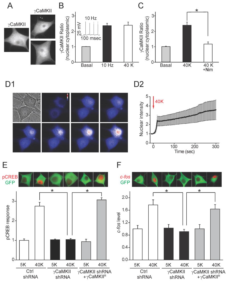
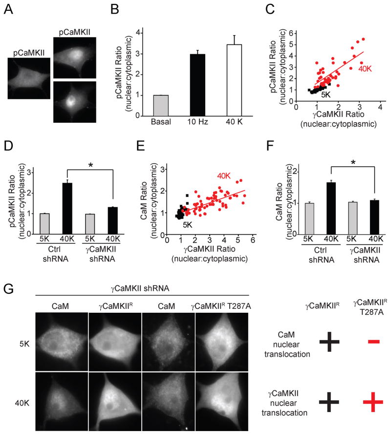
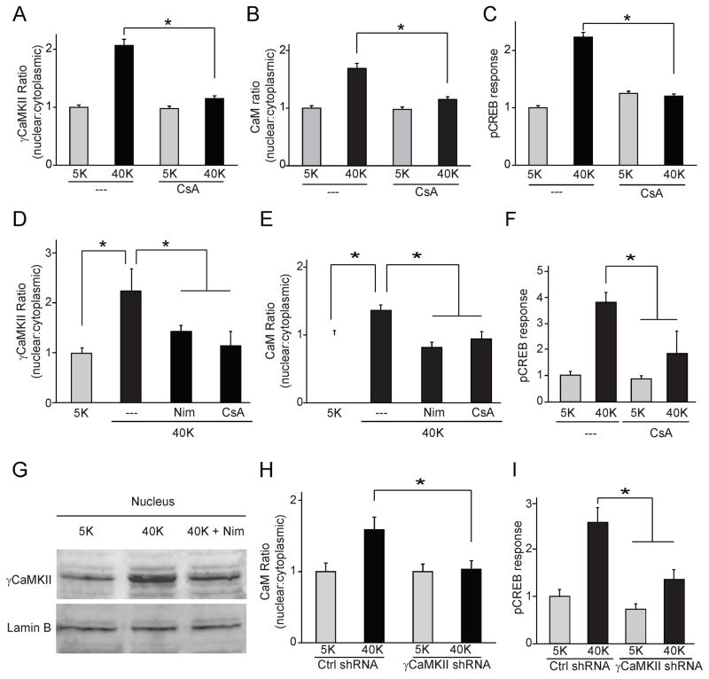
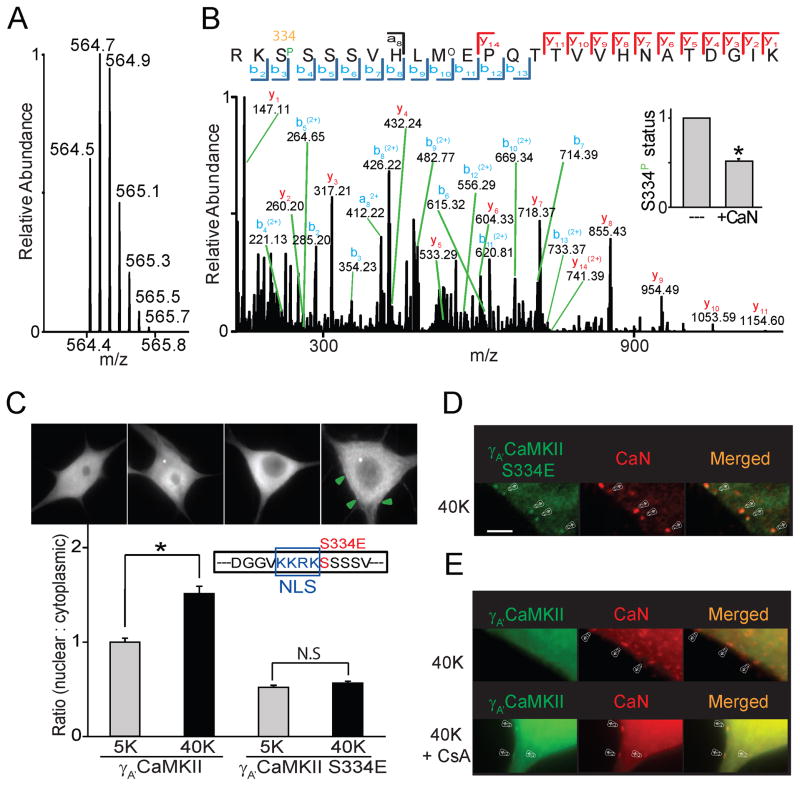
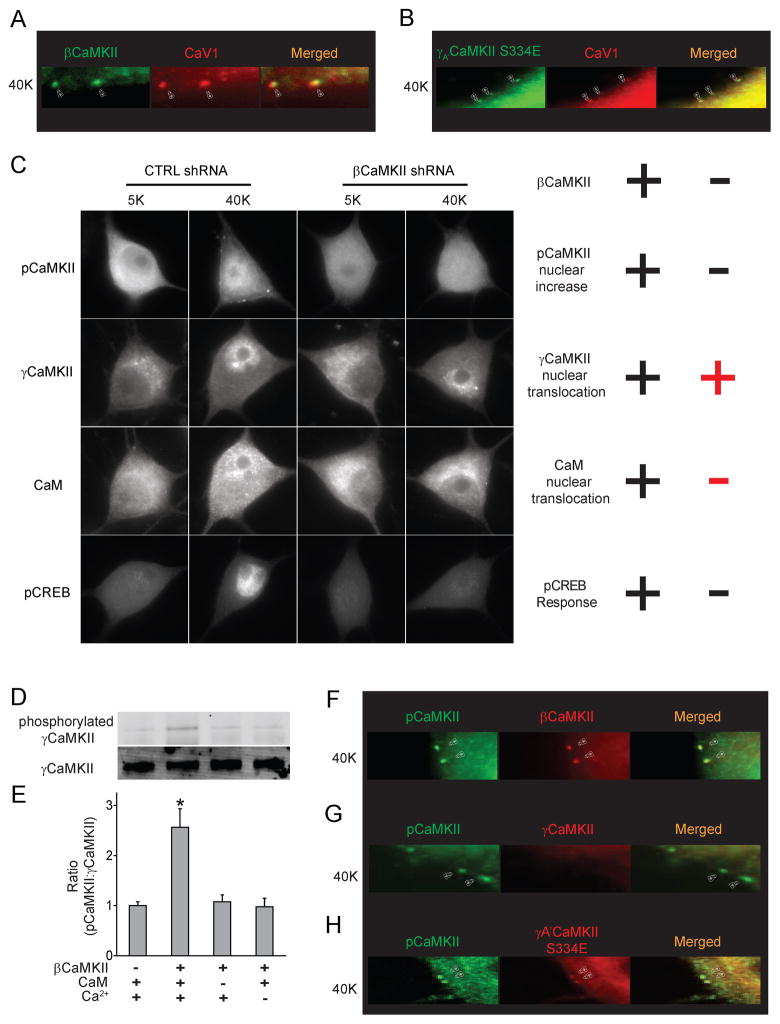
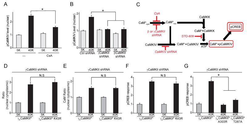
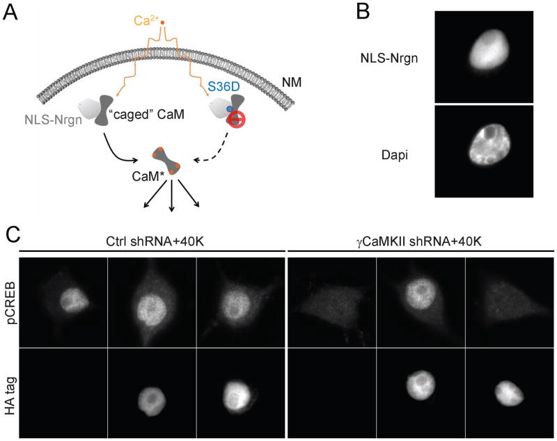
Activity-dependent CREB phosphorylation and gene expression are critical for long-term neuronal plasticity. Local signaling at CaV1 channels triggers these events, but how information is relayed onward to the nucleus remains unclear. Here, we report a mechanism that mediates long-distance communication within cells: a shuttle that transports Ca(2+)/calmodulin from the surface membrane to the nucleus. We show that the shuttle protein is γCaMKII, its phosphorylation at Thr287 by βCaMKII protects the Ca(2+)/CaM signal, and CaN triggers its nuclear translocation. Both βCaMKII and CaN act in close proximity to CaV1 channels, supporting their dominance, whereas γCaMKII operates as a carrier, not as a kinase. Upon arrival within the nucleus, Ca(2+)/CaM activates CaMKK and its substrate CaMKIV, the CREB kinase. This mechanism resolves long-standing puzzles about CaM/CaMK-dependent signaling to the nucleus. The significance of the mechanism is emphasized by dysregulation of CaV1, γCaMKII, βCaMKII, and CaN in multiple neuropsychiatric disorders.


Figures

Figure 1
Figure 1. γCaMKII translocates to the nucleus in an activity-dependent manner and is critical for E-T coupling
(A) SCG neurons were stimulated at 10 Hz for 60 s or exposed to 40 mM K+ for 300 s and stained with a γCaMKII antibody. Scale bar, 10 μm. (B) Increase in nuclear: cytoplasmic ratio ofγCaMKII intensity resulting from stimulation at 10 Hz for 60 s (solid bar) or from exposure to 40 mM K+ for 300 s (open bar). Inset, recorded action potentials evoked by 10 Hz field stimulation. (C) γCaMKII translocation triggered by 40 mM K+ stimulation was abolished by the CaV1 specific blocker Nim (10 μM). (D1) mGFP-γCaMKII translocation upon 40 mM K+ stimulation. The bright field image indicates the cell shape, and other panels show epifluorescent images (union jack format) captured at indicated time points. (D2) Nuclear mGFP intensity, before and during 40 mM K+ stimulation (onset, t=0 s, n=4). (E) SCG neurons expressing γCaMKII shRNA or non-silencing control shRNA were stimulated with 40 mM K+ for 10 s and stained for pCREB (red). GFP (green) was used to confirm expression of plasmids. The pCREB response was prevented by γCaMKII knockdown and rescued by overexpressing the shRNA-resistant γCaMKII construct γCaMKIIR (see Supplemental Fig. 1H–J for details). (F) c-fos protein levels in SCG neurons transduced as in (E) and stimulated with 40 mM K+ for 300 s, followed by 40 min incubation in normal culture medium to allow time for gene expression. γCaMKII knockdown prevented the expression of c-fos, which was rescued by overexpressed γCaMKIIR (see also Supplemental Fig. 1M). In all Figures, * denotes p<0.001, as determined by Student’s t-test. Data are represented as mean +/− SEM. See also Figure S1.
Figure 2
Figure 2. Activity-dependent nuclear pCaMKII redistribution is driven by γCaMKII/CaM translocation
(A) SCG neurons were stimulated at 10 Hz for 60 s or with 40 mM K+ for 300 s and stained for phospho-Thr-286/287-CaMKII (pCaMKII). Scale bar, 10 μm. (B) Pooled data for increase in ratio of nuclear:cytoplasmic pCaMKII intensities. (C, E) Single-cell correlation of nuclear:cytoplasmic intensity ratio between pCaMKII and γCaMKII (R=0.8) or CaM and γCaMKII (R=0.7) in response to 40 mM K+, 300 s. (D, F) SCG neurons expressing γCaMKII shRNA or non-silencing control shRNA stimulated as in (C). Increased nuclear:cytoplasmic ratios for pCaMKII and CaM were prevented by γCaMKII knockdown. (G) With endogenous γCaMKII knocked down, shRNA-resistant γCaMKIIR or γCaMKIIR T287A were overexpressed in SCG neurons. Upon stimulation as in (C), both γCaMKIIR and γCaMKIIR T287A translocated to the nucleus; however, only γCaMKIIR, not γCaMKIIR T287A, rescued CaM translocation. “+” and “−“ indicate a significant or insignificant change respectively (see Supplemental Fig. 2 I, J for details). See also Figure S2.
Figure 3
Figure 3. CaN is necessary for γCaMKII/CaM translocation and pCREB response in SCG and cortical neurons
(A, B) SCG neurons stimulated with 40 mM K+ for 300 s in absence or presence of calcineurin-specific inhibitor cyclosporin A (CsA, 50 nm). Translocation of both γCaMKII (A) and CaM (B) prevented by exposure to CsA. (C) pCREB response upon 40 mM K+ stimulation for 10 s was prevented by CsA. (D, E) Cultured cortical neurons were either mock-stimulated with 5 mM K+ or stimulated with 40 mM K+ for 60 s and stained for γCaMKII or CaM (costaining with αCaMKII antibody to label excitatory neurons). CaV1 inhibitor Nim (10 μm) or CaN inhibitor CsA (50 nm) prevented translocation ofγCaMKII and CaM. (F) Cortical neurons treated as in (C), in additional presence of specific MEK inhibitor PD98059 (50 μm), were stained for pCREB. pCREB response inhibited by CsA. (G) Nuclei isolated from cultured cortical neurons and subjected to Western blot analysis to probe for γCaMKII. 40 mM K+ stimulation of intact cells induced an increase of nuclear γCaMKII that was prevented by Nim (N=7). Lamin B is a nuclear marker. (H) Cortical neurons transfected with either γCaMKII shRNA or non-silencing control shRNA, stimulated as in (D) and stained for CaM. γCaMKII knockdown prevented CaM translocation. (I) Cortical neurons expressing either γCaMKII shRNA or a non-silencing control shRNA. Stimulated pCREB response prevented by γCaMKII knockdown. See also Figure S3.
Figure 4
Figure 4. γCAMKII is dephosphorylated by CaN near the cell surface to drive nuclear translocation
(A) Full scan mass spectrum of the quintuply charged ion (m/z 564.4773) for the phosphopeptide RKS(Phospho)SSSVHLM(oxidation)EPQTTVVHNATDGIK from γA′CaMKII. (B) Tandem mass spectrometry (MS/MS) of the phosphopeptide RKS(Phospho)SSSVHLM(Oxidation)EPQTTVVHNATDGIK. MS/MS was performed on the quintuply charged ion (m/z 564.4773) by using higher energy collision induced dissociation. The sequence of the peptide as well as the site of phosphorylation were confidently identified based on the matched b- and y-ion series indicated by the spectrum annotations. Note that all b-ions except the b2 ion correspond to fragments after neutral loss of phosphoric acid. The bar graph (insert) indicates that the phosphorylation status of S334 (marked by the orange square) was attenuated to ~50% of the control group after adding calcineurin and CaM for 30 min. N = 3 experiments, each done in triplicate. (C) SCG neurons expressing γA′CaMKII or γA′CaMKII S334E stimulated with 40 mM K+ for 300 s; γA′CaMKII but not γA′CaMKII S334E translocated to the nucleus upon stimulation. Puncta of γA′CaMKII S334E formed on the cell surface upon stimulation marked by green arrows. Scale bar, 10 μm. Inset, position of S334E point mutation, just carboxyl to the nuclear localization sequence (NLS). (D, E) Representative images of SCG neurons overexpressing γA′CaMKII S334E or γA′CaMKII, stimulated with 40 mM K+ for 60 s. Arrowheads, sites where γA′CaMKII S334E puncta colocalize with CaN (D) or γA′CaMKII puncta colocalize with CaN in presence of CaN inhibitor CsA (lower panel) (E). No γA′CAMKII puncta formed without CaN inhibitor (upper panel) (E). Scale bar, 5 μm. See also Figure S4.
Figure 5
Figure 5. Phosphorylation of γCaMKII by βCaMKII: dispensable for γCaMKII translocation but required for CaM translocation and CREB phosphorylation
(A, B) Arrowheads, sites where endogenous βCaMKII (A) or overexpressed γA′CaMKII S334E (B) colocalize with puncta of CaV1.3 channels upon 40 mM K+ stimulation for 60 s. Scale bar, 1 μm. (C) SCG neurons expressing βCaMKII shRNA or non-silencing control shRNA were stimulated with 40 mM K+ for 300 s and stained for pCaMKII (first row), co-stained for γCaMKII and CaM (second and third row), stained for pCREB (fourth row). Scale bar, 10 μm. βCaMKII knockdown prevented the increase in nuclear pCaMKII levels (−), CaM translocation (−) and pCREB response (−), but did not affect γCaMKII translocation (+). “+” and “−” indicate a significant or insignificant change respectively (see Supplemental Fig. 5A–D for details). (D–E) HA-tagged γA′CaMKII K43R was concentrated and immobilized with agarose beads coated with HA antibody. After a 10-s exposure to 25μM ATP, with or without the presence of 44 μg/ml βCaMKII, 1 mM CaCl2 and 1 μM CaM, the immobilized γA′CaMKII K43R was eluted from the beads and collected for western blot analysis. Phosphorylation state and amount of γA′CaMKII K43R detected by specific antibodies against pCaMKII and γCaMKII (D). A specific antibody to βCaMKII was used to confirm that the pCaMKII was not caused by βCaMKII accumulation on the beads. (E) Ratio of pCaMKII: γCaMKII increased significantly in the presence of βCaMKII (n=7), but only if both CaM and Ca2+ were present (n=4). (F-H) Arrowheads indicate sites where endogenous βCAMKII (F) and overexpressed γA′CaMKII S334E (G), but not γCaMKII (H), colocalize with pCaMKII puncta upon 40 mM K+ stimulation for 60 s. Scale bar, 1 μm. See also Figure S5.
Figure 6
Figure 6. Activation of nuclear CaMK cascade and distinct roles of γCaMKII and CaM in driving CREB phosphorylation
(A) SCG neurons stimulated with 40 mM K+ for 10 s in the absence or presence of CaN inhibitor CsA (50 nm). Increase in nuclear pCaMKIV levels inhibited by CsA. (B) SCG neurons expressing γCaMKII shRNA, βCaMKII shRNA or nonsilencing control shRNA were stimulated with 40 mM K+ for 10 s and stained for pCaMKIV. Knockdown of γCaMKII or βCaMKII prevented the activation of CaMKIV. (C) Schema showing free Ca2+/CaM (denoted CaM*) delivered by γCaMKII activating the CaMK cascade comprised of CaMKK and CaMKIV. Prevention of critical steps by applying different inhibitors (CsA, STO-609) or shRNA (against βCaMKII, γCaMKII or CaMKIV) indicated in red; rectangles show monitored variables (see Supplemental Fig. 6 for details). (D–F) SCG neurons expressing γCaMKII shRNA with γCaMKII shRNA-resistant γA′CaMKIIR or γA′CaMKIIR K43R were stimulated with 40 mM K+ for 300 s (D, E) or 10 s (F). Both γA′CaMKIIR and γA′CaMKIIR K43R can translocate to the nucleus upon the stimulation (D), and support CaM translocation (E) and pCREB response (F). (G) SCG neurons expressing γCaMKII shRNA with γCaMKII shRNA resistant γA′CaMKIIR A303R or γA′CaMKIIR T287A were stimulated as in (F). Neither γA′CaMKIIR A303R nor γA′CaMKIIR T287A rescued the pCREB response. See also Figure S6.
Figure 7
Figure 7. Nuclear delivery of Ca2+/CaM is sufficient to drive CREB phosphorylation
(A) Testing the sufficiency of Ca2+/CaM with a nuclear-localized, caged CaM. (B) HA-tagged NLS-Nrgn or NLS-Nrgn S36D successfully targeted to the nucleus. Scale bar, 10 μm. (C) SCG neurons expressing γCaMKII shRNA or non-silencing control shRNA, stimulated with 40 mM K+ for 10 s. NLS-Nrgn but not NLS-Nrgn S36D was able to restore the pCREB response, consistent respectively with the ability or inability to harbor CaM for release upon nuclear Ca2+ elevation. Under basal conditions, overexpressing NLS-Nrgn or NLS-Nrgn S36D did not affect pCREB levels (see Supplemental Fig. 7A, B for details). See also Figure S7.

Comment in

References

    1. Bachs O, Agell N, Carafoli E. Calmodulin and calmodulin-binding proteins in the nucleus. Cell calcium. 1994;16:289–296. - PubMed
    1. Bartsch D, Casadio A, Karl KA, Serodio P, Kandel ER. CREB1 encodes a nuclear activator, a repressor, and a cytoplasmic modulator that form a regulatory unit critical for long-term facilitation. Cell. 1998;95:211–223. - PubMed
    1. Baudier J, Deloulme JC, Van Dorsselaer A, Black D, Matthes HW. Purification and characterization of a brain-specific protein kinase C substrate, neurogranin (p17) J Biol Chem. 1991;266:229–237. - PubMed
    1. Bayer KU, Lohler J, Schulman H, Harbers K. Developmental expression of the CaM kinase II isoforms: ubiquitous gamma- and delta-CaM kinase II are the early isoforms and most abundant in the developing nervous system. Brain Res Mol Brain Res. 1999;70:147–154. - PubMed
    1. Berridge MJ. Calcium signalling remodelling and disease. Biochem Soc Trans. 2012;40:297–309. - PubMed

Publication types

MeSH terms

Substances