. 2014 Oct 9;159(2):346-57.
doi: 10.1016/j.cell.2014.09.024.

Repair of a DNA-protein crosslink by replication-coupled proteolysis

Affiliations

Repair of a DNA-protein crosslink by replication-coupled proteolysis

Julien P Duxin et al. Cell. .

Abstract

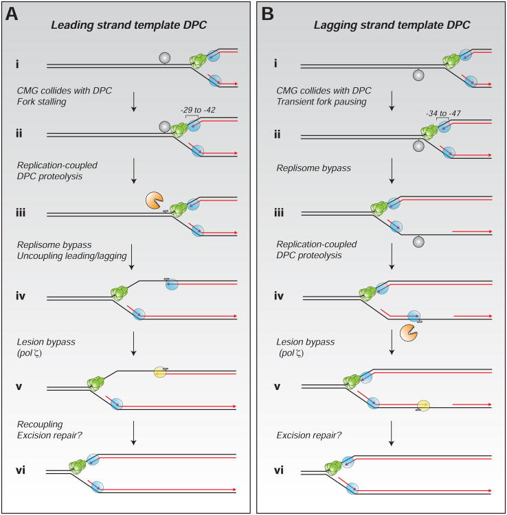
DNA-protein crosslinks (DPCs) are caused by environmental, endogenous, and chemotherapeutic agents and pose a severe threat to genome stability. We use Xenopus egg extracts to recapitulate DPC repair in vitro and show that this process is coupled to DNA replication. A DPC on the leading strand template arrests the replisome by stalling the CMG helicase. The DPC is then degraded on DNA, yielding a peptide-DNA adduct that is bypassed by CMG. The leading strand subsequently resumes synthesis, stalls again at the adduct, and then progresses past the adduct using DNA polymerase ζ. A DPC on the lagging strand template only transiently stalls the replisome, but it too is degraded, allowing Okazaki fragment bypass. Our experiments describe a versatile, proteolysis-based mechanism of S phase DPC repair that avoids replication fork collapse.


Figures

Figure 1
Figure 1. Replication of a plasmid containing a site-specific DPC
(A) Schematic of pDPCTop. (B) pCTRL and pDPCTop were replicated in egg extract in the presence of [α-32P]dATP. Samples were treated with Proteinase K (ProtK), as indicated, and analyzed by agarose gel electrophoresis. OC, open circular; SC, supercoiled. (C) pCTRL and pDPCTop were replicated, digested with FspI, and analyzed by 2-D gel electrophoresis. The lower cartoon illustrates relevant DNA intermediates. Arrows, see main text. (D) Model for how replication of pDPC initially yields 50% open circular (OC) and 50% supercoiled (SC) products.
Figure 2
Figure 2. Multistep bypass of a DPC
Depiction of nascent leading strands generated after NcoI digestion of DPCTop (A) and pDPCBot (B). Extension products were monitored with NcoI and AatII digestion. Green hexamer, rightward CMG helicase; N, sequencing primer. (C) pCTRL, pDPCTop, and pDPCBot replication intermediates were digested with NcoI and AatII (upper panel) or NcoI (lower panel), and separated on a denaturing polyacrylamide gel alongside a sequencing ladder. Nascent strands generated by the rightward fork are indicated in brackets. For lagging strand identification, see Figure legend S2C. Red and grey arrows, extension products of the top (Top) and bottom (Bot) strands (upper panel). Black arrows, location of the DPC in pDPCTop and pDPCBot (bottom panel).
Figure 3
Figure 3. The DPC is degraded on DNA
(A) Strategy to detect adducted parental strands. (B) pCTRL and pDPCTop were replicated without [α-32P]dATP and supplemented with Geminin (Gem.) where indicated. DNA was phenol-chloroform extracted, digested with NcoI and NdeI, and separated on a denaturing polyacrylamide gel. The top strand was detected by Southern blotting. Where indicated, samples were treated with Proteinase K (ProtK) prior to phenol-chloroform extraction. Plasmids containing an intact DPC were lost in the organic layer during phenol-chloroform extraction. This was the case in all samples where no replication occurred (lanes 3, 5, and 6), suggesting DPC destruction is replication-dependent. (C) Schematic of assay to monitor DPC proteolysis. b, biotin. (D) pDPCTop containing biotinylated M.HpaII was replicated with [α-32P]dATP. Input DNA (lower autoradiograph) and DNA co-precipitated with streptavidin (upper autoradiograph) were treated with ProtK and analyzed on an agarose gel. OC, open circular; SC, supercoiled. (E) DNA recovered in the pull-down (panel D) was quantified and normalized to the input, and compared to the appearance of Top strand extension product, as in lanes 8-14 of Figure 2C (primary data not shown).
Figure 4
Figure 4. DNA pol ζ depletion inhibits DPC bypass
(A) Mock-depleted and Rev7-depleted egg extracts were blotted with Rev7 and MCM7 antibodies. (B) The extracts from (A) were used for replication of pDPCBot as in Figure 1B (+ProtK). (C) Samples from (B) were analyzed as in Figure 2C.
Figure 5
Figure 5. DPC bypass by a single replication fork
Depiction of nascent leading strand intermediates generated when DPC-LTop (A) or pDPC-LBot (B) are replicated in the presence of LacI (blue spheres) and then digested with AatII. Digestion with FspI and AatII yields Top extension products that are 4 nt shorter than Bot extension products. In the presence of LacI, Top extension products correspond to leading strands, and Bot extension products to lagging strands. (C) pCTRL-L, pDPC-LTop, and pDPC-LBot were replicated in the presence of buffer or LacI. Samples were digested with FspI and AatII (upper panel) or AatII alone (lower panel), and analyzed on a denaturing polyacrylamide gel. Colored brackets, nascent strands generated by the leftward fork (lower panel). Arrows, extension products of the Top and Bot strands (upper panel). (D) pDPC-LTop and pDPC-LBot prepared with biotinylated M.HpaII were replicated in the presence of LacI. Samples were analyzed as in Figure 3D. (E) The pull-down recovery of pDPC-LTop in (D) was graphed alongside the -29 to -42 leading strand arrest quantified from Figure S5D. Values were normalized to the maximum peak value. (F) The pull-down recovery of pDPC-LBot in (D) was graphed alongside the -34 to -47 leading strand arrest quantified from Figure S5D.
Figure 6
Figure 6. DPC processing is essential for efficient replicative DPC bypass
(A) pDPC-LTop and pDPC-LBot were replicated in the presence of LacI and buffer (+Buffer), 13 μM Ub-VS (+Ub-VS) or 13 μM Ub-VS and 80 μM ubiquitin (+Ub-VS +Ub.). Samples were analyzed as in Figure 3D. (B) Samples from (A) were analyzed as in Figure 5C. (C) Quantification of the -29 to -42 species of pDPC-LTop from panel (B). Values were normalized to the maximum peak value of the buffer control. (D) -34 to -47 species of pDPC-LBot from panel (B) were quantified as in (C).
Figure 7
Figure 7. Model for DPC repair in Xenopus egg extracts
Repair of a DPC on the leading (A) or lagging strand templates (B). See Discussion for details. Black lines, parental DNA; red lines, nascent strands; in green, CMG helicase; in blue, replicative polymerases; in yellow, TLS polymerase; in grey, DPC.

References

    1. Arias EE, Walter JC. Replication-dependent destruction of Cdt1 limits DNA replication to a single round per cell cycle in Xenopus egg extracts. Genes Dev. 2005;19:114–126. - PMC - PubMed
    1. Baker DJ, Wuenschell G, Xia L, Termini J, Bates SE, Riggs AD, O'Connor TR. Nucleotide Excision Repair Eliminates Unique DNA-Protein Cross-links from Mammalian Cells. Journal of Biological Chemistry. 2007;282:22592–22604. - PubMed
    1. Barker S, Weinfeld M, Murray D. DNA-protein crosslinks: their induction, repair, and biological consequences. Mutat Res. 2005;589:111–135. - PubMed
    1. Chen L, MacMillan AM, Chang W, Ezaz-Nikpay K, Lane WS, Verdine GL. Direct identification of the active-site nucleophile in a DNA (cytosine-5)-methyltransferase. Biochemistry. 1991;30:11018–11025. - PubMed
    1. Chvalova K, Brabec V, Kasparkova J. Mechanism of the formation of DNA-protein cross-links by antitumor cisplatin. Nucleic Acids Res. 2007;35:1812–1821. - PMC - PubMed

Publication types