. 2014 Oct 9;159(2):428-39.
doi: 10.1016/j.cell.2014.09.040.

Generation of functional human pancreatic β cells in vitro

Affiliations

Generation of functional human pancreatic β cells in vitro

Felicia W Pagliuca et al. Cell. .

Abstract

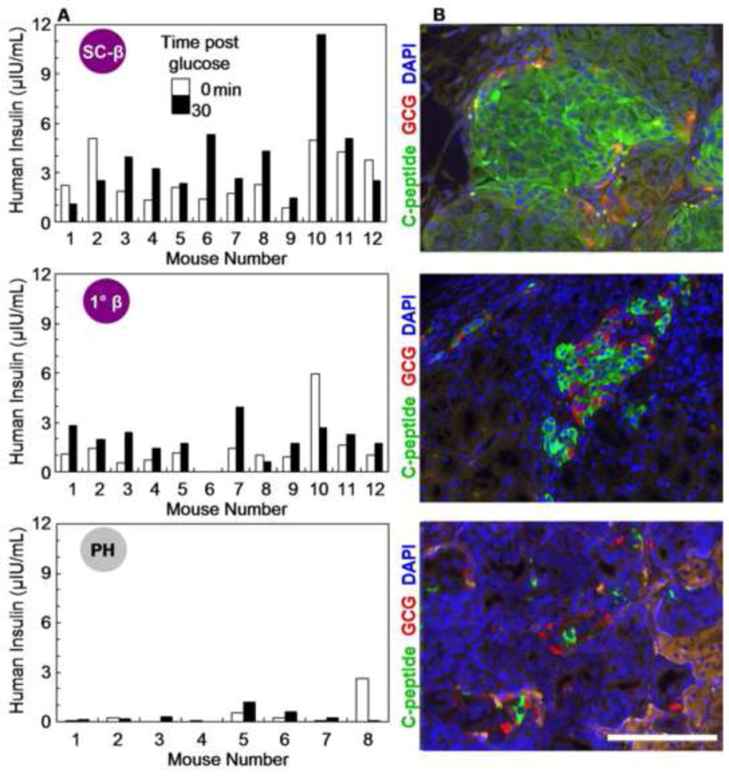
The generation of insulin-producing pancreatic β cells from stem cells in vitro would provide an unprecedented cell source for drug discovery and cell transplantation therapy in diabetes. However, insulin-producing cells previously generated from human pluripotent stem cells (hPSC) lack many functional characteristics of bona fide β cells. Here, we report a scalable differentiation protocol that can generate hundreds of millions of glucose-responsive β cells from hPSC in vitro. These stem-cell-derived β cells (SC-β) express markers found in mature β cells, flux Ca(2+) in response to glucose, package insulin into secretory granules, and secrete quantities of insulin comparable to adult β cells in response to multiple sequential glucose challenges in vitro. Furthermore, these cells secrete human insulin into the serum of mice shortly after transplantation in a glucose-regulated manner, and transplantation of these cells ameliorates hyperglycemia in diabetic mice.


Figures

Figure 1
Figure 1. SC-β cells generated in vitro secrete insulin in response to multiple sequential high glucose challenges like primary human β cells
(A) Schematic of directed differentiation from hPSC into INS+ cells via new or previously published control differentiations. (B–D) Representative ELISA measurements of secreted human insulin from HUES8 SC-β cells (B), PH cells (C), and primary β (1°β) cells (D) challenged sequentially with 2, 20, 2, 20, 2, and 20mM glucose, with a 30-min incubation for each concentration (see Methods). After sequential low/high glucose challenges, cells were depolarized with 30mM KCl. (E–G) Box and whisker plots of secreted human insulin from different biological batches of HUES8 (open circles) and hiPSC SC-β (black circles) cells (E; n=12), biological batches of PH cells (F; n=5), and primary β cells (G; n=4). Each circle is the average value for all sequential challenges with 2mM or 20mM glucose in a batch. Insulin secretion at 20mM ranged 0.23–2.7 μIU/103 cells for SC-β cells and 1.5–4.5 μIU/103 cells for human islets, and the stimulation index ranged 0.4–4.1 for SC-β cells and 0.6–4.8 for primary adult. The thick horizontal line indicates the median. See also Figures S1 and S2A and Table S1. * p<0.05 when comparing insulin secretion at 20mM vs. 2mM with paired t-test Act A=Activin A; CHIR=CHIR99021, a GSK3α/β inhibitor; KGF= keratinocyte growth factor or FGF family member 7; RA=Retinoic Acid; SANT1=sonic hedgehog pathway antagonist; LDN=LDN193189, a BMP type 1 receptor inhibitor; PdbU=Phorbol 12,13-dibutyrate, a protein kinase C activator; Alk5i=Alk5 receptor inhibitor II; T3=triiodothyronine, a thyroid hormone; XXI=γ-secretase inhibitor; Betacellulin=EGF family member
Figure 2
Figure 2. SC-β cells flux cytosolic Ca2+ in response to multiple sequential high glucose challenges like primary human β cells
(A) Schematic of population level and single cell level detection of cytosolic Ca2+ using Fluo-4 AM staining. Population level measurements were taken on individual whole clusters (marked by large red circle in the schematic), and individual cells within intact clusters (marked by small red circles) were analyzed for single cell analysis. (B) Representative population measurements of dynamic normalized Fluo-4 fluorescence intensity for HUES8 SC-β cells, primary β cells, and PH cells challenged sequentially with 2, 20, 2, 20, 2, and 20mM glucose and 30mM KCl. The x-axis represents time (in s). (C) Fluorescence images of Fluo-4 AM staining used in single cell analysis. (D) Representative images showing single cells that responded to 3 (yellow), 2 (orange), 1 (blue), and 0 (red) glucose challenges. (E) Quantification of the frequency of SC-β cells (n=156), primary β cells (n=114), and PH cells (n=138) that responded to 20mM glucose. Scale bar = 100 μm
Figure 3
Figure 3. SC-β cells express human β cell markers at protein and gene expression level
(A) Representative immunohistochemistry (IHC) of HUES8 SC-β cells, primary β cells, and PH cells stained for C-peptide (green) and NKX6-1 (red). (B) Representative IHC of cells stained for C-peptide (green) and PDX1 (red). (C) Representative IHC of cells stained for C-peptide (green) and glucagon (red) with the corresponding DAPI stain (blue). Enlarged insets in A, B and C shows staining for transcription factors (NKX6-1 and PDX1) is nuclear and cytoplasmic for C-peptide, except for the PH cells. (D) Representative flow cytometry dot plots and population percentages of cells stained for C-peptide and NKX6-1. AU = arbitrary units. (E) Hierarchal clustering based on all genes measured by transcriptional microarray of undifferentiated HUES8, PH cells, fetal β cells, and adult primary β cells sorted for INS (data from Hrvatin et al. 2014), and SC-β cells sorted for INS and NKX6-1. (F) Heat map of the 100 genes with the most variance across all samples. All images were taken with a Zeiss LSM 710 confocal microscope. See also Figures S2B–C, S3, and S4. CP=C-peptide, SST=somatostatin, GCG=glucagon Scale bar=100 μm
Figure 4
Figure 4. SC-β cell insulin granules are structurally similar to primary human β cell granules
(A) Electron microscopy images of granules within sectioned cells, highlighting representative crystallized insulin granules (red), early insulin granules (yellow), and mixed endocrine granules (blue) found in HUES8 SC-β cells, primary β cells, and PH cells. Scale bar=500 nm. (B) Higher magnification images of granules highlighted in (A). Scale bar=500 nm. (C) Box and whisker plot of the number of insulin and early insulin granules per cell. The cross indicates mean and the thick horizontal line indicates median. n=24 cells from 2 batches of differentiation for HUES8 SC-β cells, and n=30 cells from 2 donors of primary human β cells. PH cells are not shown because no mature insulin or early insulin granules were observed. (D) Electron microscopy images of cells labeled with immunogold staining showing granules that contain insulin (smaller 5 nm black dots) and/or glucagon (larger 15 nm black dots). Representative immunogold particles are highlighted with red arrows (insulin) and blue arrows (glucagon). Scale bar=100 nm.
Figure 5
Figure 5. Transplanted SC-β cells function rapidly in vivo
(A) ELISA measurements of human insulin from the serum of individual mice transplanted with comparable numbers of HUES8 SC-β cells (5×10⁶ cells), primary human β cells (500–1000 IEQ), or PH cells (5×10⁶ cells). Measurements were taken before (white bars) and 30 min after (black bars) a glucose injection of mice 2 wk post-transplantation. (B) Representative IHC of cells 2 wk post-transplantation in (A) stained with C-peptide (green) and glucagon (GCG; red) to confirm presence of graft. All images were taken with a Olympus IX51 microscope. See also Figure S5 and Tables S2 and S3. Scale bar=100 μm
Figure 6
Figure 6. Transplanted SC-β cells rapidly ameliorates hyperglycemia in diabetic mice
(A) Fasting blood glucose measurements of progressively diabetic NRG-Akita mice transplanted with HUES8 SC-β cells (5×10⁶ cells; open circles; n=6) or PH cells (5×10⁶ cells; closed square; n=6 for 0, 18, and 28 d and n=4 for 53, 84 and 112 d). Glucose measurements were saturated at 600mg/dL. *p<0.05 comparing the 2 cell groups on the same d with unpaired t-test. Data presented as mean±SEM. (B) Blood glucose measurements of mice that have been transplanted 137 d prior with HUES8 SC-β cells (n=5) or PH cells (n=1) and a separate independent cohort of mice transplanted 34 d prior with human islets (4000 IEQ; closed triangle; n=6). Note: It was not possible to coordinate receipt of human cadaveric islets and the production of SC-β cells. So the data in Figure 6B come from a different animal cohort. One mouse that received HUES8 SC-β cells died after a blood draw taken in (C), and 5 mice that received PH cells died over the duration of the observation period. Data presented as mean±SEM. (C) ELISA measurements of human insulin from the serum of individual mice transplanted with HUES8 SC-β cells (n=6) or PH cells (n=2). Mice were fasted for 16 hr and then blood glucose was measured before (white bars) and 30 min after (black bars) a glucose injection of mice, 126 d post-transplantation. p<0.05 for transplanted HUES8 SC-β cells insulin concentration 30 min vs. 0 min after glucose injection with paired t-test.

Comment in

  • Diabetes: β cells at last.
    Mfopou JK, Bouwens L. Mfopou JK, et al. Nat Rev Endocrinol. 2015 Jan;11(1):5-6. doi: 10.1038/nrendo.2014.200. Epub 2014 Nov 11. Nat Rev Endocrinol. 2015. PMID: 25385037 No abstract available.
  • Stem cells: Insulin-producing β cells in a dish.
    Baumann K. Baumann K. Nat Rev Mol Cell Biol. 2014 Dec;15(12):768. doi: 10.1038/nrm3907. Epub 2014 Nov 12. Nat Rev Mol Cell Biol. 2014. PMID: 25387398 No abstract available.
  • 'Diabetes: a cure at last' or just hype?
    Hitman GA. Hitman GA. Diabet Med. 2014 Dec;31(12):1479. doi: 10.1111/dme.12611. Diabet Med. 2014. PMID: 25399773 No abstract available.

References

    1. Aguayo-Mazzucato C, Zavacki AM, Marinelarena A, Hollister-Lock J, Khattabi IE, Marsili A, Weir GC, Sharma A, Larsen PR, Bonner-Weir S. Thyroid Hormone Promotes Postnatal Rat Pancreatic β-Cell Development and Glucose-Responsive Insulin Secretion Through MAFA. Diabetes. 2013;62:1569–580. - PMC - PubMed
    1. Apelqvist A, Li H, Sommer L, Beatus P, Anderson DJ, Honjo T, Hrabe de Angelis M, Lendahl U, Edlund H. Nature. 1999;400:877–881. - PubMed
    1. Bellin MD, Barton FB, Heitman A, Harmon J, Balamurugan AN, Kandaswamy R, Sutherland DE, Alejandro R, Hering BJ. Potent Induction Immunotherapy Promotes Long-Term Insulin Independence After Islet Transplantation in Type 1 Diabetes. American Journal of Transplantation. 2012;12:1576–583. - PMC - PubMed
    1. Blum B, Hrvatin SS, Schuetz C, Bonal C, Rezania A, Melton DA. Functional beta-cell maturation is marked by an increased glucose threshold and by expression of urocortin 3. Nat Biotechnol. 2012 Feb 26;30(3):261–4. 2012. - PMC - PubMed
    1. Brehm MA, Bortell R, Diiorio P, Leif J, Laning J, Cuthbert A, Yang C, Herlihy M, Burzenski L, et al. Human Immune System Development and Rejection of Human Islet Allografts in Spontaneously Diabetic NOD-Rag1null IL2r{gamma} Ins2Akita Mice. Diabetes. 2010;59:2265–2270. - PMC - PubMed

Publication types

Associated data