

Tumor necrosis factor alpha promotes the proliferation of human nucleus pulposus cells via nuclear factor-κB, c-Jun N-terminal kinase, and p38 mitogen-activated protein kinase
- PMID: 25304312
- PMCID: PMC4935371
- DOI: 10.1177/1535370214554533
Tumor necrosis factor alpha promotes the proliferation of human nucleus pulposus cells via nuclear factor-κB, c-Jun N-terminal kinase, and p38 mitogen-activated protein kinase
Abstract
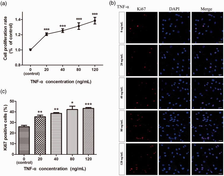
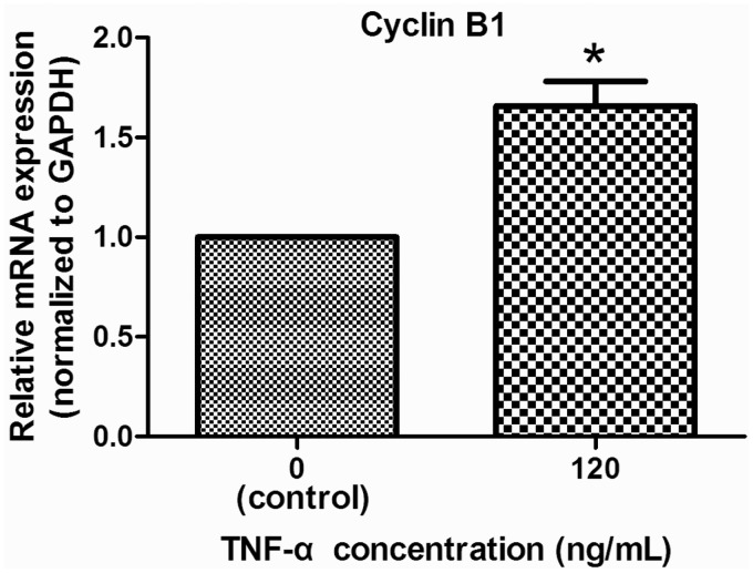
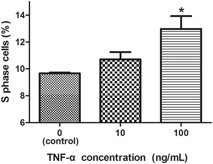
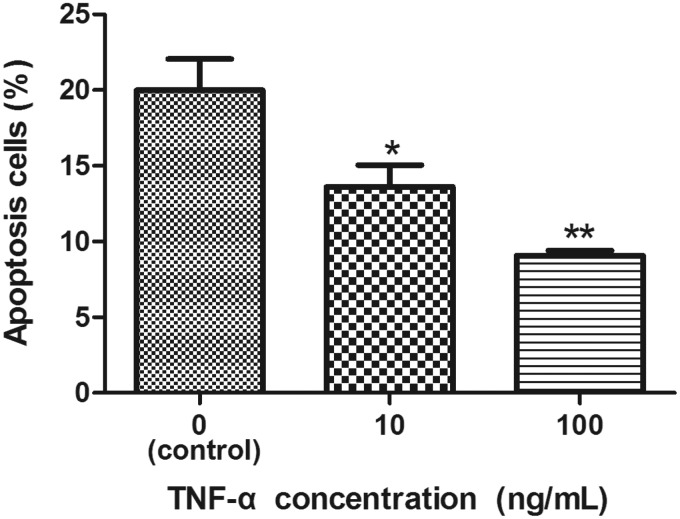
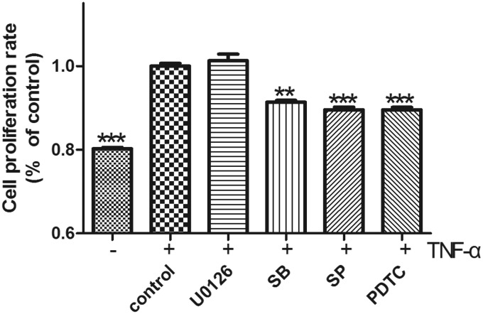
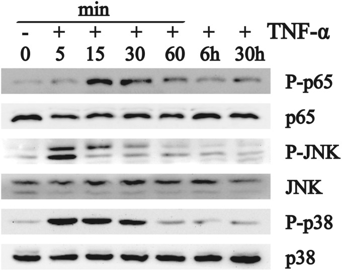
Although tumor necrosis factor alpha (TNF-α) is known to play a critical role in intervertebral disc (IVD) degeneration, the effect of TNF-α on nucleus pulposus (NP) cells has not yet been elucidated. The aim of this study was to explore the effect of TNF-α on proliferation of human NP cells. NP cells were treated with different concentrations of TNF-α. Cell proliferation was determined by cell counting kit-8 (CCK-8) analysis and Ki67 immunofluorescence staining, and expression of cyclin B1 was studied by quantitative real-time RT-PCR. Cell cycle was measured by flow cytometry and cell apoptosis was analyzed using an Annexin V-fluorescein isothiocyanate (FITC) & propidium iodide (PI) apoptosis detection kit. To identify the mechanism by which TNF-α induced proliferation of NP cells, selective inhibitors of major signaling pathways were used and Western blotting was carried out. Treatment with TNF-α increased cell viability (as determined by CCK-8 analysis) and expression of cyclin B1 and the number of Ki67-positive and S-phase NP cells, indicating enhancement of proliferation. Consistent with this, NP cell apoptosis was suppressed by TNF-α treatment. Moreover, inhibition of NF-κB, c-Jun N-terminal kinase (JNK), and p38 mitogen-activated protein kinase (MAPK) blocked TNF-α-stimulated proliferation of NP cells. In conclusion, the current findings suggest that the effect of TNF-α on IVD degeneration involves promotion of the proliferation of human NP cells via the NF-κB, JNK, and p38 MAPK pathways.
Keywords: Tumor necrosis factor alpha; intervertebral disc degeneration; mitogen-activated protein kinase; nuclear factor-κB; nucleus pulposus cell; proliferation.
© 2014 by the Society for Experimental Biology and Medicine.
Figures
References
-
- Takahashi H, Suguro T, Okazima Y, Motegi M, Okada Y, Kakiuchi T. Inflammatory cytokines in the herniated disc of the lumbar spine. Spine 1996; 21: 218–24. - PubMed
-
- Doita M, Kanatani T, Ozaki T, Matsui N, Kurosaka M, Yoshiya S. Influence of macrophage infiltration of herniated disc tissue on the production of matrix metalloproteinases leading to disc resorption. Spine 2001; 26: 1522–7. - PubMed
-
- Weiler C, Nerlich AG, Bachmeier BE, Boos N. Expression and distribution of tumor necrosis factor alpha in human lumbar intervertebral discs: a study in surgical specimen and autopsy controls. Spine 2005; 30: 44–53. - PubMed
-
- Miyamoto H, Saura R, Harada T, Doita M, Mizuno K. The role of cyclooxygenase-2 and inflammatory cytokines in pain induction of herniated lumbar intervertebral disc. Kobe J Med Sci 2000; 46: 13–28. - PubMed
-
- Rand N, Reichert F, Floman Y, Rotshenker S. Murine nucleus pulposus-derived cells secrete interleukins-1-β,-6, and-10 and granulocyte-macrophage colony-stimulating factor in cell culture. Spine 1997; 22: 2598–601. - PubMed
MeSH terms
Substances
LinkOut - more resources
Full Text Sources
Other Literature Sources
Research Materials
Miscellaneous