

Development of PEA-15 using a potent non-viral vector for therapeutic application in breast cancer
- PMID: 25304382
- PMCID: PMC4571276
- DOI: 10.1016/j.canlet.2014.09.033
Development of PEA-15 using a potent non-viral vector for therapeutic application in breast cancer
Abstract
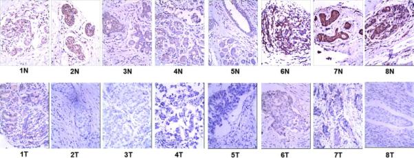
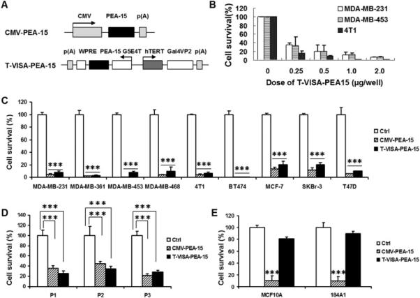
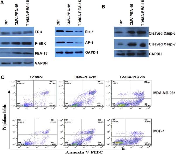
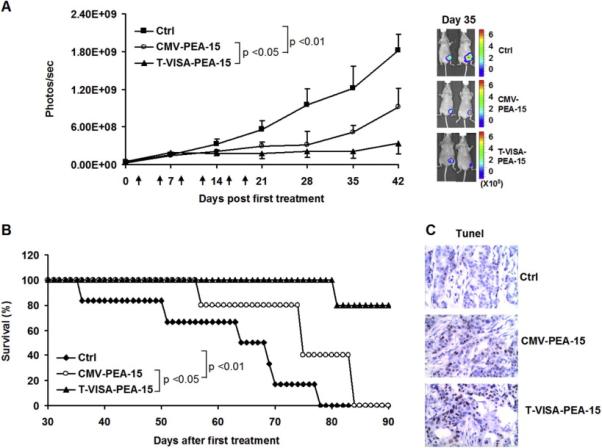
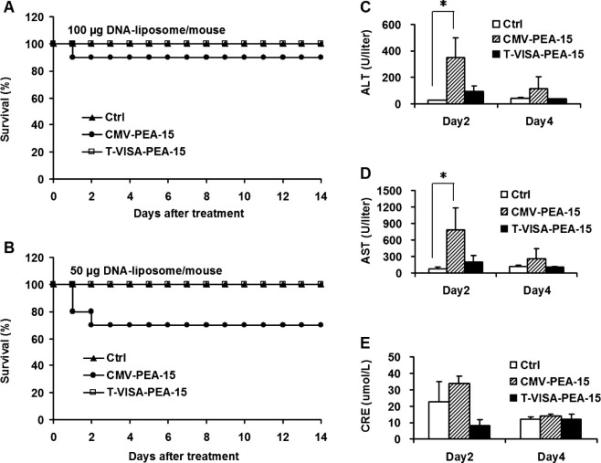
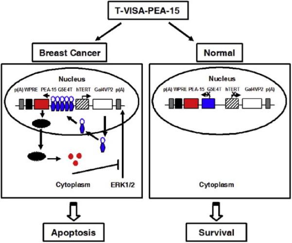
Advanced breast cancer requires systemic treatment, therefore developing an efficient and safe strategy is urgently needed. To ensure the success of target therapy, we have developed a breast cancer-specific construct (T-VISA) composed of the human telomerase reverse transcriptase (hTERT; T) promoter and a versatile transgene amplification vector VISA (VP16-GAL4-WPRE integrated systemic amplifier) to target PEA-15 (phosphoprotein enriched in astrocytes) in advanced breast tumors. PEA-15 contains a death effector domain that sequesters extracellular signal-regulated kinase (ERK) in the cytoplasm, thereby inhibiting cell proliferation and inducing apoptosis. T-VISA-PEA-15 was found to be highly specific, selectively express PEA-15 in breast cancer cells, and induce cancer-cell killing in vitro and in vivo without affecting normal cells. Moreover, intravenous treatment with T-VISA-PEA-15 coupled with liposome nanoparticles attenuated tumor growth and prolonged survival in mice bearing advanced breast tumors. Importantly, there was virtually no severe toxicity when PEA-15 is expressed by our T-VISA system compared with cytomegalovirus (CMV) promoter. Thus, our findings demonstrate an effective cancer-targeted therapy that is worthy of development in clinical trials eradicating advanced breast cancer.
Keywords: Breast cancer; PEA-15; T-VISA system; Target therapy.
Copyright © 2014 Elsevier Ireland Ltd. All rights reserved.
Figures
Similar articles
-
Targeted expression of BikDD eliminates breast cancer with virtually no toxicity in noninvasive imaging models.Mol Cancer Ther. 2012 Sep;11(9):1915-24. doi: 10.1158/1535-7163.MCT-12-0191. Epub 2012 Jul 2. Mol Cancer Ther. 2012. PMID: 22752427
-
PEA-15 inhibits tumorigenesis in an MDA-MB-468 triple-negative breast cancer xenograft model through increased cytoplasmic localization of activated extracellular signal-regulated kinase.Clin Cancer Res. 2010 Mar 15;16(6):1802-11. doi: 10.1158/1078-0432.CCR-09-1456. Epub 2010 Mar 9. Clin Cancer Res. 2010. PMID: 20215547
-
Targeted endostatin-cytosine deaminase fusion gene therapy plus 5-fluorocytosine suppresses ovarian tumor growth.Oncogene. 2013 Feb 28;32(9):1082-90. doi: 10.1038/onc.2012.134. Epub 2012 May 7. Oncogene. 2013. PMID: 22562248
-
Cancer-specific gene therapy.Adv Genet. 2005;54:235-55. doi: 10.1016/S0065-2660(05)54010-0. Adv Genet. 2005. PMID: 16096014 Review.
-
Phosphoprotein enriched in astrocytes (PEA)-15: a potential therapeutic target in multiple disease states.Pharmacol Ther. 2014 Sep;143(3):265-74. doi: 10.1016/j.pharmthera.2014.03.006. Epub 2014 Mar 20. Pharmacol Ther. 2014. PMID: 24657708 Free PMC article. Review.
Cited by
-
Metformin mediates induction of miR-708 to inhibit self-renewal and chemoresistance of breast cancer stem cells through targeting CD47.J Cell Mol Med. 2019 Sep;23(9):5994-6004. doi: 10.1111/jcmm.14462. Epub 2019 Jul 5. J Cell Mol Med. 2019. PMID: 31273952 Free PMC article.
-
Nanoparticle Delivery of miR-34a Eradicates Long-term-cultured Breast Cancer Stem Cells via Targeting C22ORF28 Directly.Theranostics. 2017 Oct 17;7(19):4805-4824. doi: 10.7150/thno.20771. eCollection 2017. Theranostics. 2017. PMID: 29187905 Free PMC article.
-
Non-Phosphorylatable PEA-15 Sensitises SKOV-3 Ovarian Cancer Cells to Cisplatin.Cells. 2020 Feb 24;9(2):515. doi: 10.3390/cells9020515. Cells. 2020. PMID: 32102425 Free PMC article.
-
On the Quest of Cellular Functions of PEA-15 and the Therapeutic Opportunities.Pharmaceuticals (Basel). 2015 Jul 31;8(3):455-73. doi: 10.3390/ph8030455. Pharmaceuticals (Basel). 2015. PMID: 26263999 Free PMC article. Review.
-
Phosphorylation of PED/PEA-15 at Ser116 and phosphorylation of p27 at Thr187 indicates a poor prognosis in hepatocellular carcinoma.Oncol Lett. 2021 Mar;21(3):177. doi: 10.3892/ol.2021.12438. Epub 2021 Jan 6. Oncol Lett. 2021. PMID: 33574916 Free PMC article.
References
-
- Siegel R, Ma J, Zou Z, Jemal A. Cancer statistics, 2014. CA: a cancer journal for clinicians. 2014;64:9–29. - PubMed
-
- Dou S, Yao YD, Yang XZ, Sun TM, Mao CQ, Song EW, Wang J. Anti-Her2 single-chain antibody mediated DNMTs-siRNA delivery for targeted breast cancer therapy. Journal of controlled release : official journal of the Controlled Release Society. 2012;161:875–883. - PubMed
-
- Yao YD, Sun TM, Huang SY, Dou S, Lin L, Chen JN, Ruan JB, Mao CQ, Yu FY, Zeng MS, Zang JY, Liu Q, Su FX, Zhang P, Lieberman J, Wang J, Song E. Targeted delivery of PLK1-siRNA by ScFv suppresses Her2+ breast cancer growth and metastasis. Science translational medicine. 2012;4:130–148. - PubMed
-
- Xie X, Guo J, Kong Y, Xie GX, Li L, Lv N, Xiao X, Tang J, Wang X, Liu P, Yang M, Xie Z, Wei W, Spencer DM, Xie X. Targeted expression of Escherichia coli purine nucleoside phosphorylase and Fludara(R) for prostate cancer therapy. The journal of gene medicine. 2011;13:680–691. - PubMed
Publication types
MeSH terms
Substances
Grants and funding
LinkOut - more resources
Full Text Sources
Other Literature Sources
Medical
Miscellaneous
