. 2015 Feb;64(2):149-59.
doi: 10.1007/s00262-014-1618-8. Epub 2014 Oct 11.

Patients with pancreatic adenocarcinoma exhibit elevated levels of myeloid-derived suppressor cells upon progression of disease

Affiliations

Patients with pancreatic adenocarcinoma exhibit elevated levels of myeloid-derived suppressor cells upon progression of disease

Joseph Markowitz et al. Cancer Immunol Immunother. 2015 Feb.

Abstract

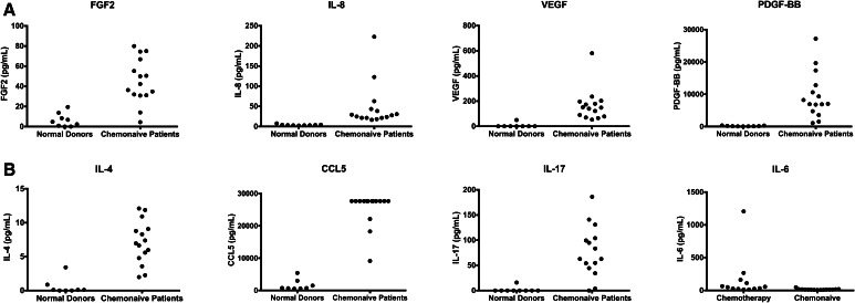
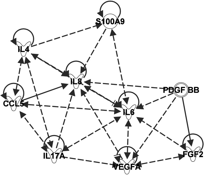
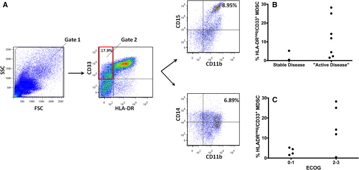
Elevated levels of myeloid-derived suppressor cells (MDSCs) induced by tumor-derived factors are associated with inhibition of immune responses in patients with gastrointestinal malignancies. We hypothesized that pro-MDSC cytokines and levels of MDSC in the peripheral blood would be elevated in pancreatic adenocarcinoma patients with progressive disease. Peripheral blood mononuclear cells (PBMCs) were isolated from 16 pancreatic cancer patients undergoing chemotherapy and phenotyped for MDSC using a five antigen panel (CD33, HLA-DR, CD11b, CD14, CD15). Patients with stable disease had significantly lower MDSC levels in the peripheral blood than those with progressive disease (1.41 ± 1.12 vs. 5.14 ± 4.58 %, p = 0.013, Wilcoxon test). A cutoff of 2.5 % MDSC identified patients with progressive disease. Patients with ECOG performance status ≥2 had a weaker association with increased levels of MDSC. Plasma was obtained from 15 chemonaive patients, 13 patients undergoing chemotherapy and 9 normal donors. Increases in the levels of pro-MDSC cytokines were observed for pancreatic cancer patients versus controls, and the pro-MDSC cytokine IL-6 was increased in those patients undergoing chemotherapy. This study suggests that MDSC in peripheral blood may be a predictive biomarker of chemotherapy failure in pancreatic cancer patients.


Conflict of interest statement

The authors declare that they have no conflict of interest.

Figures

Fig. 1
Fig. 1
a Levels of cytokines are increased in the plasma of chemonaive patients versus normal donors. The MDSC levels in 15 patients with pancreatic cancer naive to chemotherapy were compared to nine normal donors. Five patients had stage IIB disease, five patients had stage III disease, and five patients had stage IV disease. b Levels of IL-6 are increased in the plasma of pancreatic adenocarcinoma patients undergoing chemotherapy as compared to chemonaive patients; 15 chemonaive patients (population described in a) were compared to 13 patients undergoing chemotherapy. In the patients undergoing chemotherapy, three patients had stage II disease, two patients had stage III disease, and eight patients had stage IV disease
Fig. 2
Fig. 2
Pathway analysis of cytokines determined to be up-regulated in plasma collected from pancreatic adenocarcinoma patients. The pathway map was generated by adding the cytokines increased in the plasma of untreated pancreatic adenocarcinoma and those cytokines that were found to be increased in chemotherapy-treated pancreatic adenocarcinoma versus chemonaive into a new pathway map in IPA and have IPA determine the relationships. S100A9 was then added to the map given the importance of S100A9 in the myeloid chemotaxis pathway, and IPA was utilized to predict the relationships between the cytokines and S100A9. IL-6 is a central regulator of MDSC biology and pathway analysis using Ingenuity Pathway Analysis software confirmed this finding. In addition, the protein S100A9, although not part of the BioPlex® analysis, appears to be important in signaling in pancreatic adenocarcinoma patients. Dashed line arrows signify indirect relationships. Solid lines with arrows represent direct relationships. A solid line parallel to the arrow signifies inhibition. Proteins that regulate themselves have a solid line pointed to themselves (e.g., S100A9, IL-4, CCL5 (RANTES), IL-6). The proteins have different shapes based on their categorization in IPA. Cytokines have a Y shape. S100A9 and PDGF-BB are not placed in any one particular group which is why they have circular shapes
Fig. 3
Fig. 3
a Representative scatter plots for a pancreatic adenocarcinoma patient. In the left most panel, the scatter plot (side scatter vs. forward scatter) is presented. The center plot shows the staining for CD33 and HLA-DR. The scatter plot was then created for CD15 versus CD11b to determine the percentage of granulocytic MDSC. CD14 versus CD11b was plotted for monocytic MDSC. b HLA-DRneg/CD33+ percentages in PBMCs measured as a function of whether they had stable (n = 2), or “active disease” (n = 7) in a retrospective study of nine patients with pancreatic adenocarcinoma. Active disease was defined by progressive disease on chemotherapy or a new diagnosis of pancreatic adenocarcinoma. One patient had stage II disease, one patient had stage III disease, and seven patients had stage IV disease. This analysis led to the hypothesis that the MDSC percentage increases with progressive disease that was tested with a prospectively collected cohort of patients. c HLA-DRneg/CD33+ percentages in PBMCs obtained from peripheral blood measured as a function of ECOG performance status. An ECOG score of 0–1 indicates good performance status (n = 4) and a score of 2–3 indicates poor performance status (n = 5)
Fig. 4
Fig. 4
a PBMCs were measured using two markers for cell populations negative for HLA-DR and positive for CD33. Patients were categorized according to whether they had stable (n = 5) or progressive disease (n = 11). A HLA-DRnegCD33+ percentage greater than 2.5 % is suggestive of progressive disease. Of these 16 patients, 1 patient had stage I disease, 2 patients had stage II disease, four patients had stage III disease, and 9 patients had stage IV disease. b Measurement of HLA-DRnegCD33+CD14negCD15neg in the same patient populations suggests a percentage greater than 0.5 % lineage-negative MDSC is suggestive of progressive disease. c Percentages of HLA-DRnegCD33+ cells were measured as a function of ECOG performance status. Worse performance status is associated with increasing numbers of MDSC. Patients with ECOG ≥ 2 (n = 5) tended to have higher levels of MDSC

References

    1. Siegel R, Naishadham D, Jemal A. Cancer statistics, 2013. CA Cancer J Clin. 2013;63(1):11–30. doi: 10.3322/caac.21166. - DOI - PubMed
    1. Conroy T, Desseigne F, Ychou M, Bouche O, Guimbaud R, Becouarn Y, Adenis A, Raoul JL, Gourgou-Bourgade S, de la Fouchardiere C, Bennouna J, Bachet JB, Khemissa-Akouz F, Pere-Verge D, Delbaldo C, Assenat E, Chauffert B, Michel P, Montoto-Grillot C, Ducreux M. FOLFIRINOX versus gemcitabine for metastatic pancreatic cancer. N Engl J Med. 2011;364(19):1817–1825. doi: 10.1056/NEJMoa1011923. - DOI - PubMed
    1. Kim R. FOLFIRINOX: a new standard treatment for advanced pancreatic cancer? Lancet Oncol. 2011;12(1):8–9. doi: 10.1016/S1470-2045(10)70237-0. - DOI - PubMed
    1. Ying JE, Zhu LM, Liu BX. Developments in metastatic pancreatic cancer: Is gemcitabine still the standard? World J Gastroenterol. 2012;18(8):736–745. doi: 10.3748/wjg.v18.i8.736. - DOI - PMC - PubMed
    1. Kothari N, Saif MW, Kim R. First-line treatment for advanced pancreatic cancer. JOP. 2013;14(2):129–132. - PubMed

Publication types

MeSH terms