. 2015 Jan 1;308(1):L1-10.
doi: 10.1152/ajplung.00222.2014. Epub 2014 Oct 10.

Force maintenance and myosin filament assembly regulated by Rho-kinase in airway smooth muscle

Affiliations

Force maintenance and myosin filament assembly regulated by Rho-kinase in airway smooth muscle

Bo Lan et al. Am J Physiol Lung Cell Mol Physiol. .

Abstract

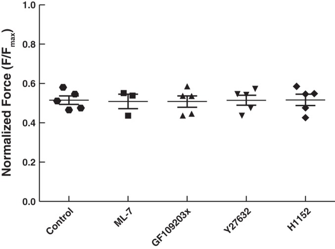
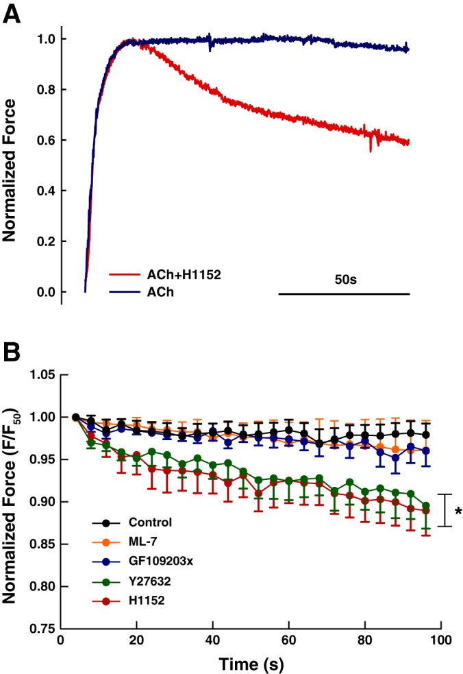
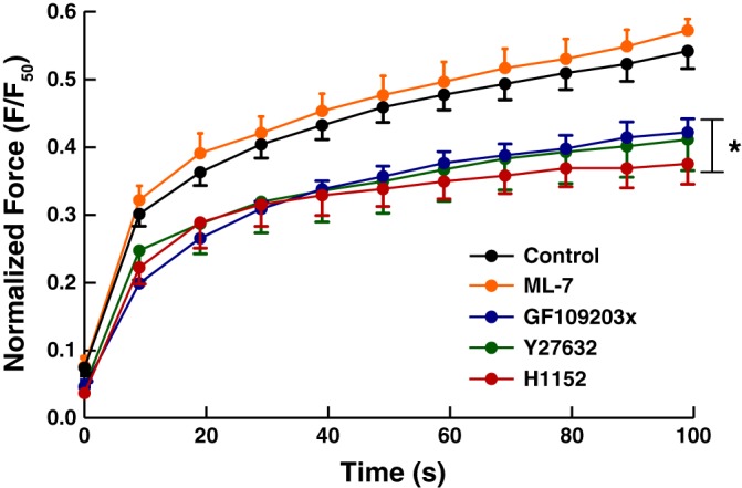
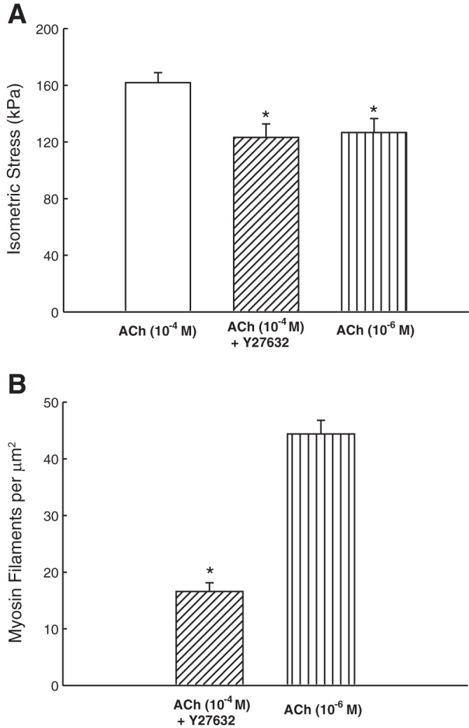
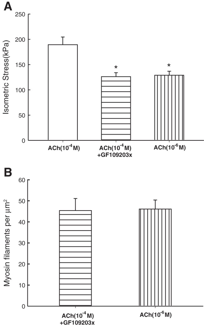
Smooth muscle contraction can be divided into two phases: the initial contraction determines the amount of developed force and the second phase determines how well the force is maintained. The initial phase is primarily due to activation of actomyosin interaction and is relatively well understood, whereas the second phase remains poorly understood. Force maintenance in the sustained phase can be disrupted by strains applied to the muscle; the strain causes actomyosin cross-bridges to detach and also the cytoskeletal structure to disassemble in a process known as fluidization, for which the underlying mechanism is largely unknown. In the present study we investigated the ability of airway smooth muscle to maintain force after the initial phase of contraction. Specifically, we examined the roles of Rho-kinase and protein kinase C (PKC) in force maintenance. We found that for the same degree of initial force inhibition, Rho-kinase substantially reduced the muscle's ability to sustain force under static conditions, whereas inhibition of PKC had a minimal effect on sustaining force. Under oscillatory strain, Rho-kinase inhibition caused further decline in force, but again, PKC inhibition had a minimal effect. We also found that Rho-kinase inhibition led to a decrease in the myosin filament mass in the muscle cells, suggesting that one of the functions of Rho-kinase is to stabilize myosin filaments. The results also suggest that dissolution of myosin filaments may be one of the mechanisms underlying the phenomenon of fluidization. These findings can shed light on the mechanism underlying deep inspiration induced bronchodilation.

Keywords: asthma; bronchoprotection; muscle mechanics; ultrastructure.


Figures

Fig. 1.
Fig. 1.
Protocol for determining maintenance of peak force (during stretch) and force recovery after length oscillation. In experiments where enzyme inhibitors were used, the muscle was preincubated with an inhibitor for a predetermined period of time (see text) before commencement of this protocol.
Fig. 2.
Fig. 2.
Force generation from all groups matched to 50% Fmax (maximal contraction induced by ACh 10−4 M). For the control group, a lower dose of Ach (within the range of 10−7 to 10−5 M) was used to stimulate the muscle to match 50% Fmax; for the 4 groups with inhibitors, the force generation was induced by 10−4 M Ach with the inhibitors. There is no statistically significant difference among the groups (1-way ANOVA, P = 0.992). The average Fmax values are 215.5 ± 37.4 kPa (n = 5) for the control group, 207.9 ± 39.3 kPa (n = 3) for the ML-7 group, 232.5 ± 23.7 kPa (n = 5) for the GF109023x group, 189.7 ± 17.6 kPa (n = 4) for the Y27632 group, and 198.1 ± 23.6 kPa (n = 5) for the H1152 group.
Fig. 3.
Fig. 3.
A: representative force records for ACh (10−6 M) stimulation with and without H1152 (3 μM). B: force maintenance over a 100-s period. Both H1152 and Y27632 groups are significantly different compared with control, ML-7, and GF109203x groups (2-way ANOVA, *P < 0.0001). The two Rho-kinase inhibitors are not different from each other (2-way ANOVA, P = 0.1846.)
Fig. 4.
Fig. 4.
Decline of peak force due to oscillation. Force record in the absence of oscillation (Fig. 2B) was subtracted from force record with the same inhibitor in the presence of oscillation. Note that in the presence of oscillatory strain only the peak force during each stretch was recorded. The Y27632 and the H1152 groups are significantly different from control, ML-7, and GF109203x (2-way ANOVA, *P < 0.0001). Incubation with GF109203x and ML-7 had no significant effect on force response during length oscillation compared with control (2-way ANOVA, P = 0.294 and P = 0.8280, respectively).
Fig. 5.
Fig. 5.
Force recovery after length oscillation. The ML-7 and the control group are not significantly different (2-way ANOVA, P = 0.1245). The H1152, Y27632, and GF109203x groups are significantly different from control and ML-7 group (2-way ANOVA, *P < 0.0001), The GF109203x group is not different from the H1152 and Y27632 groups (2-way ANOVA, P = 0.1305 and P = 0.8451, respectively), and the two Rho-kinase inhibitors are not different from each other (2-way ANOVA, P = 0.2708). Force measured at time points 60, 70, 80, 90, and 100 s in the H1152 group are significantly different compared with the control; for the Y27632 group, only force measured at 100 s is significantly different from the control (Bonferroni post tests, P < 0.001).
Fig. 6.
Fig. 6.
Examples of electron micrographs showing cross-sections of trachealis. A: partially ACh-activated trachealis. B: magnified area from A. C: ACh-activated trachealis in the presence Rho-kinase inhibitor (Y27632) and force-matched to A. D: magnified area from C. E: partially ACh-activated trachealis. F: magnified area from E. G: ACh-activated trachealis in the presence PKC inhibitor (GF109203x) and force-matched to E. H: magnified area from G. M, mitochondrion; SR, sarcoplasmic reticulum; DB, dense body; DP, dense plaque; Cav, caveola. Unlabeled arrows in the magnified areas point to myosin filaments surrounded by actin filaments; double arrows point to intermediate filaments. Myosin filaments have irregular cross-sectional profile and an average diameter of 15 nm, larger than that of actin filaments (6 nm) and that of intermediate filaments (10 nm, circular profile), but smaller than dense bodies. Scale bar, 0.5 μm.
Fig. 6.
Fig. 6.
Examples of electron micrographs showing cross-sections of trachealis. A: partially ACh-activated trachealis. B: magnified area from A. C: ACh-activated trachealis in the presence Rho-kinase inhibitor (Y27632) and force-matched to A. D: magnified area from C. E: partially ACh-activated trachealis. F: magnified area from E. G: ACh-activated trachealis in the presence PKC inhibitor (GF109203x) and force-matched to E. H: magnified area from G. M, mitochondrion; SR, sarcoplasmic reticulum; DB, dense body; DP, dense plaque; Cav, caveola. Unlabeled arrows in the magnified areas point to myosin filaments surrounded by actin filaments; double arrows point to intermediate filaments. Myosin filaments have irregular cross-sectional profile and an average diameter of 15 nm, larger than that of actin filaments (6 nm) and that of intermediate filaments (10 nm, circular profile), but smaller than dense bodies. Scale bar, 0.5 μm.
Fig. 7.
Fig. 7.
A: maximal (plateau) stress produced by fully activated (ACh, 10−4 M), Y27632 inhibited (10−4 M ACh + 3 μM Y27632), and partially activated (ACh, in the range of 10−6.5–10−6 M) muscles. *Significant difference (1-way ANOVA, P < 0.05) from fully activated force. B: myosin filament density in Y27632 inhibited and partially activated muscles. *Significant difference (1-way ANOVA, P < 0.001).
Fig. 8.
Fig. 8.
A: maximal (plateau) stress produced by fully activated (ACh, 10−4 M), GF109203x-inhibited (10−4 M ACh + 15 μM GF109203x), and partially activated (ACh, in the range of 10−7–10−5 M) muscles. *Significant difference (1-way ANOVA, P < 0.05) from fully activated force. B: there was no significant difference in the myosin filament densities in GF109203x and its ACh-activated control (1-way ANOVA, P = 0.924).
Fig. 9.
Fig. 9.
Simulation results using the mathematical model to test the role of myosin filament length distribution on force maintenance in sustained contraction. Here we mimicked the experimental protocol both for a standard filament length distribution (solid line), and an altered distribution with a greater fraction of shorter filaments (dashed line) to observe the alteration in force maintained during the oscillation protocol (see materials and methods). The favorable agreement with the experimental data (c.f. Fig. 3) suggests that changes to the filament length distribution may play an important role in regulating this force response.
Fig. 10.
Fig. 10.
Schematic illustration of contractile units containing myosin filament fragments. It is assumed that one of the functions of Rho-kinase is to stabilize myosin filaments. With Rho-kinase inhibitor (+), myosin filaments breakdown into shorter fragments; some of the fragments become invisible to EM so that the myosin filament density would appear to decrease (see text for more description).

References

    1. An SS, Bai TR, Bates JH, Black JL, Brown RH, Brusasco V, Chitano P, Deng L, Dowell M, Eidelman DH, Fabry B, Fairbank NJ, Ford LE, Fredberg JJ, Gerthoffer WT, Gilbert SH, Gosens R, Gunst SJ, Halayko AJ, Ingram RH, Irvin CG, James AL, Janssen LJ, King GG, Knight DA, Lauzon AM, Lakser OJ, Ludwig MS, Lutchen KR, Maksym GN, Martin JG, Mauad T, McParland BE, Mijailovich SM, Mitchell HW, Mitchell RW, Mitzner W, Murphy TM, Paré PD, Pellegrino R, Sanderson MJ, Schellenberg RR, Seow CY, Silveira PS, Smith PG, Solway J, Stephens NL, Sterk PJ, Stewart AG, Tang DD, Tepper RS, Tran T, Wang L. Airway smooth muscle dynamics: a common pathway of airway obstruction in asthma. Eur Respir J 29: 834–860, 2007. - PMC - PubMed
    1. Brook BS. Emergence of airway smooth muscle mechanical behavior through dynamic reorganization of contractile units and force transmission pathways. J Appl Physiol 116: 980–997, 2014. - PMC - PubMed
    1. Chen C, Krishnan R, Zhou E, Ramachandran A, Tambe D, Rajendran K, Adam RM, Deng L, Fredberg JJ. Fluidization and resolidification of the human bladder smooth muscle cell in response to transient stretch. PLos One 5: e12035, 2010. - PMC - PubMed
    1. Chin LY, Bossé Y, Pascoe C, Hackett TL, Seow CY, Paré PD. Mechanical properties of asthmatic airway smooth muscle. Eur Respir J 40(1): 45–54, 2012. - PubMed
    1. Craig R, Smith R, Kendrick-Jones J. Light-chain phosphorylation controls the conformation of vertebrate non-muscle and smooth muscle myosin molecules. Nature 302: 436–439, 1983. - PubMed

Publication types