. 2014 Nov;20(11):1279-88.
doi: 10.1038/nm.3654. Epub 2014 Oct 12.

Osteoblast-derived WNT16 represses osteoclastogenesis and prevents cortical bone fragility fractures

Affiliations

Osteoblast-derived WNT16 represses osteoclastogenesis and prevents cortical bone fragility fractures

Sofia Movérare-Skrtic et al. Nat Med. 2014 Nov.

Abstract

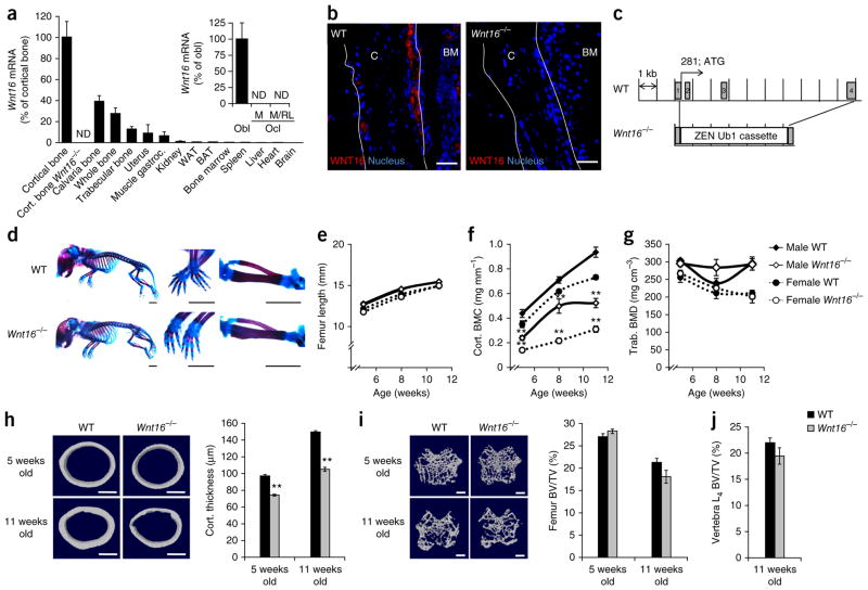
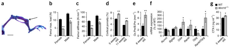
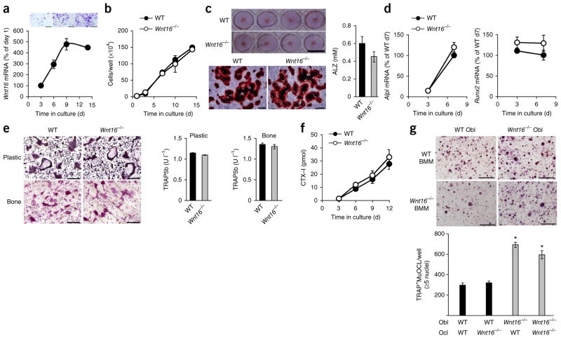
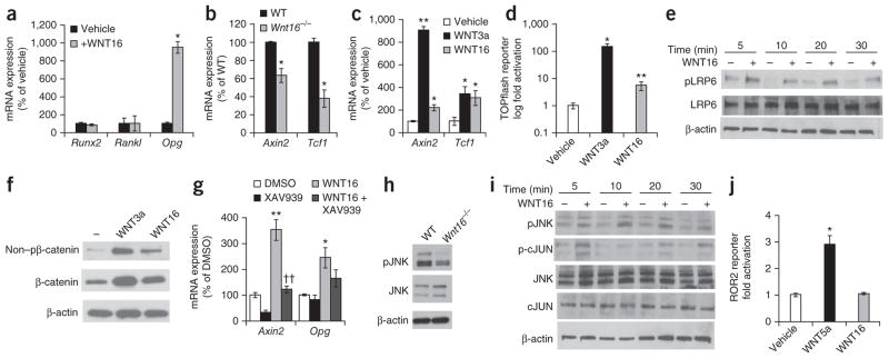
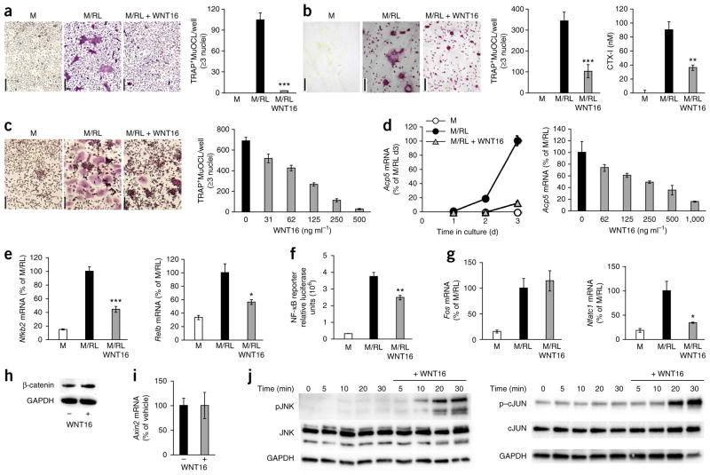
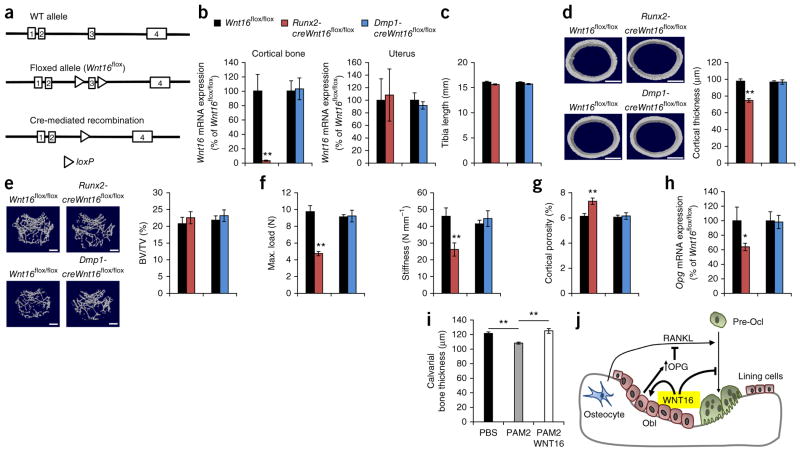
The WNT16 locus is a major determinant of cortical bone thickness and nonvertebral fracture risk in humans. The disability, mortality and costs caused by osteoporosis-induced nonvertebral fractures are enormous. We demonstrate here that Wnt16-deficient mice develop spontaneous fractures as a result of low cortical thickness and high cortical porosity. In contrast, trabecular bone volume is not altered in these mice. Mechanistic studies revealed that WNT16 is osteoblast derived and inhibits human and mouse osteoclastogenesis both directly by acting on osteoclast progenitors and indirectly by increasing expression of osteoprotegerin (Opg) in osteoblasts. The signaling pathway activated by WNT16 in osteoclast progenitors is noncanonical, whereas the pathway activated in osteoblasts is both canonical and noncanonical. Conditional Wnt16 inactivation revealed that osteoblast-lineage cells are the principal source of WNT16, and its targeted deletion in osteoblasts increases fracture susceptibility. Thus, osteoblast-derived WNT16 is a previously unreported key regulator of osteoclastogenesis and fracture susceptibility. These findings open new avenues for the specific prevention or treatment of nonvertebral fractures, a substantial unmet medical need.


Conflict of interest statement

COMPETING FINANCIAL INTERESTS

The authors declare no competing financial interests.

Figures

Figure 1
Figure 1
Wnt16−/− mice have reduced cortical but not trabecular bone mass. (a) Wnt16 mRNA levels in different tissues. Cort., cortical; gastroc., gastrocnemius; WAT, white adipose tissue; BAT, brown adipose tissue; Obl, primary cultured osteoblasts; Ocl, primary cultured osteoclasts; M, macrophage colony-stimulating factor (M-CSF) stimulation; M/RL, M-CSF and RANKL stimulation; ND, not detectable (n = 6). (b) Immunohistochemistry analysis of WNT16 immunostaining (red) and nuclear staining (blue, DAPI) in the cortical bone (C) of WT and Wnt16−/− mice. BM, bone marrow. The white outlines indicate the cortical bone surfaces. Scale bars, 50 μm. (c) Schematic of the Wnt16 gene in the generated Wnt16−/−, exon 1-4 mice. (d) Whole-mount Alizarin red and Alcian blue staining of Wnt16−/− and WT embryos (E18.5) with close ups of the paws (middle) and tibia and fibula (right). Scale bars, 2.5 mm. (eg) Femur lengths, cortical bone mineral content (BMC) and trabecular BMD of the tibia as measured by peripheral quantitative CT (pQCT) in Wnt16−/− and WT mice at 5 (n = 9), 8 (n = 9) and 11 (female WT, n = 9; female Wnt16−/−, n = 8; male WT, n = 11; male Wnt16−/−, n = 12) weeks of age. (h,i) Cortical thickness (h) and distal metaphyseal trabecular bone volume per total volume (BV/TV; i) as measured by micro-CT in the femurs of 5- and 11-week-old female Wnt16−/− and WT mice (5 weeks old: WT, n = 9; Wnt16−/−, n = 8; 11 weeks old: n = 6). Left, representative micro-CT images of the experiment described to the right. Scale bars, 500 μm. (j) Bone volume per total volume as measured by histomorphometry of trabecular bone in the L4 vertebra of 11-week-old female Wnt16−/− (n = 6) and WT (n = 7) mice. All values are given as the mean ± s.e.m. **P < 0.01, Student’s t test compared to WT.
Figure 2
Figure 2
Spontaneous fractures as a result of several defects of cortical bone in Wnt16−/− mice. (a) Spontaneous fracture of the tibia (arrow) in a Wnt16−/− mouse. Scale bar, 500 μm. (b,c) Maximal (max.) load (b) and stiffness (c) at failure determined by three-point bending of the femur in 8-week-old Wnt16−/− mice compared to WT mice (n = 9). (d) Cortical porosity in the femur diaphysis as measured by micro-CT of female Wnt16−/− compared to WT mice (5 weeks old: WT, n = 9; Wnt16−/−, n = 8; 11 weeks old: n = 6). (e) Osteoclast surface/bone surface (Oc.Pm/B.Pm) in femur cortical bone of 8-week-old female Wnt16−/− (n = 9) compared to WT (n = 7) mice. (f) mRNA expression analyses of femur cortical bone in 8-week-old female Wnt16−/− (n = 8) and WT (n = 9) mice. Rankl/Opg ratio is the ratio of the two transcripts. (g) Serum levels of the bone resorption marker CTX-I in 5-week-old Wnt16−/− and WT mice (n = 9). *P < 0.05, **P < 0.01, Student’s t test compared to WT. All values are given as the mean ± s.e.m.
Figure 3
Figure 3
Osteoblast-derived WNT16 inhibits osteoclastogenesis. (ad) Calvarial osteoblast cultures. (a) Wnt16 mRNA expression (bottom) and staining for alkaline phosphatase (top) during differentiation of osteoblasts. Scale bars, 500 μm. d, day. (b) Proliferation of Wnt16−/− and WT osteoblasts. (c) Alizarin red staining of mineralized nodules in Wnt16−/− and WT osteoblast cultures (left) and quantification of eluted Alizarin red (ALZ, right). Scale bars, 1 cm (top); 500 μm (bottom). (d) mRNA expression of Alpl and Runx2. (e) Number of TRAP+MuOCLs and release of TRAP5b into the medium in RANKL-stimulated bone marrow cultures from Wnt16−/− and WT mice on plastic (e, top images and left graph) or bone (e, bottom images and right graph). Scale bars, 400 μm. (f) Release of CTX-I from RANKL-stimulated bone marrow cells from Wnt16−/− mice cultured on bone. (g) Cocultures of BMMs and calvarial osteoblasts from Wnt16−/− and WT mice. Representative of two separate TRAP staining carried out in triplicate (top) and quantification (bottom) of TRAP+MuOCLs in cocultures. Scale bars, 200 μm. *P < 0.05, Student’s t test compared to WT Obl and WT BMMs or WT Obl and Wnt16−/− BMMs. All values are given as the mean ± s.e.m.
Figure 4
Figure 4
WNT16 increases OPG expression and signals through both canonical and noncanonical pathways in osteoblasts. (a) mRNA expression analyses of MC3T3-E1 cells treated with WNT16 (n = 3). (b) mRNA expression analyses of cortical bone isolated from WT and Wnt16−/− mice (n = 4). (c) mRNA expression analyses of MC3T3-E1 cells treated with vehicle, WNT3a or WNT16 (n = 3). (d) TOPflash luciferase assay of MC3T3-E1 cells treated with vehicle, WNT3a or WNT16. Data are presented as a fold change normalized to Renilla compared to vehicle-treated cells. (e) Western blot of phosphorylated and total LRP6 in MC3T3-E1 cells treated with WNT16. Blots are representative of three separate experiments carried out in triplicate. (f) Nonphosphorylated levels of β-catenin (non–pβ-catenin) and β-catenin in MC3T3-E1 cells treated with vehicle, 100 ng ml−1 WNT3a or 100 ng ml−1 WNT16. (g) mRNA expression analyses of MC3T3-E1 cells treated with or without WNT16 in the presence or absence of XAV939 (n = 3). Data are presented as a fold change normalized to 18S compared to vehicle-treated cells. (h) Phosphorylated levels of JNK in calvariae isolated from 3-day-old WT and Wnt16−/− mice. (i) Western blot of phosphorylated JNK and cJUN in MC3T3-E1 cells treated with WNT16. (j) Luciferase assay of HEK293 cells stably expressing ROR2-hTRK-luciferase (luc) treated with vehicle, WNT5a or WNT16. Data are presented as a fold change normalized to vehicle-treated cells. All values are given as the mean ± s.e.m. *P < 0.05, **P < 0.01, Student’s t test compared to WT or vehicle. ††P < 0.01, Student’s t test compared to WNT16-treated cells.
Figure 5
Figure 5
WNT16 inhibits osteoclast differentiation through noncanonical WNT pathways. (a,b) RANKL-stimulated osteoclastogenesis of mouse BMMs on plastic (a, TRAP staining) and bone surface (b, TRAP staining) treated with recombinant WNT16. Scale bars, 100 μm. Numbers of TRAP+MuOCLs on plastic (a) and bone (b, left graph) and release of CTX-I from bone slices (b, right graph). (c) WNT16 dose-dependent inhibition of RANKL-stimulated osteoclastogenesis in human CD14+ monocyte cultures. Left, TRAP staining of RANKL-stimulated osteoclastogenesis with or without WNT16. Scale bars, 100 μm. Right, number of TRAP+MuOCLs per well. (d) Time-dependent (left) and dose-dependent (right) inhibition of Acp5 mRNA by WNT16 in RANKL-stimulated BMM cultures. (e) mRNA expression of Nfkb2 (left) and Relb (right) in RANKL-stimulated BMM cultures with or without WNT16. (f) NF-κB luciferase gene reporter assay in BMMs cultured in M, M/RL or M/RL plus WNT16. (g) mRNA expression of Fos (left) and Nfatc1 (right) in RANKL-stimulated BMM cultures with or without WNT16. (h) Lack of effect by WNT16 on β-catenin protein levels as measured by western blot analysis. GAPDH, glyceraldehyde 3-phosphate dehydrogenase. (i) Axin2 mRNA levels in BMMs treated with WNT16. (j) Levels of phosphorylated JNK (left) and phosphorylated cJUN (right) measured by western blot in BMMs cultured with or without WNT16. Blots are representative of three separate experiments carried out in triplicate. All values are shown as the mean ± s.e.m. (n = 4). *P < 0.05, **P < 0.01, ***P < 0.005, Student’s t test compared to no WNT16.
Figure 6
Figure 6
Osteoblasts are the principal source of WNT16, with an impact on cortical bone and fracture susceptibility. (a) Schematic presentation of the WT allele, the floxed allele and the Cre-mediated recombination of Wnt16. (b) Wnt16 mRNA expression in the cortical bone (left) and uterus (right) of 5-week-old Runx2-creWnt16flox/flox (n = 9) and Dmp1-creWnt16flox/flox (n = 8) mice. (ce) Bone length (c), femur diaphyseal cortical thickness (d) and trabecular bone volume to total volume (e) in the distal femur metaphysis of Runx2-creWnt16flox/flox and Dmp1-creWnt16flox/flox mice compared to littermate controls (Wnt16flox/flox; n = 8; 5 weeks old). Cortical thickness and trabecular BV/TV were analyzed using micro-CT. (d,e, left) Representative micro-CT images of the experiment described to the right. Scale bars, 500 μm. (f) Three-point bending of femur diaphysis demonstrating maximal load (left) and stiffness at failure (right) in female Runx2-creWnt16flox/flox and Dmp1-creWnt16flox/flox mice compared to littermate controls (Wnt16flox/flox; n = 8; 5 weeks old). (g) Micro-CT analysis of cortical porosity in the femurs of female Runx2-creWnt16flox/flox and Dmp1-creWnt16flox/flox mice compared to littermate controls (Wnt16flox/flox; n = 8; 5 weeks old). (h) Opg mRNA expression in cortical bone of 5-week-old Runx2-creWnt16flox/flox (n = 9) and Dmp1-creWnt16flox/flox (n = 8) mice. (i) WNT16 treatment of PAM2-induced calvarial bone loss after 5 d as measured by micro-CT (PBS, n = 6; PAM2, n = 6; PAM2 + WNT16, n = 5). (j) Proposed role of WNT16 in cortical bone. WNT16 is osteoblast derived and inhibits human and mouse osteoclastogenesis both directly by acting on osteoclast progenitors and indirectly by increasing OPG expression in osteoblasts. *P < 0.05, **P < 0.01, Student’s t test compared to control. All values are given as the mean ± s.e.m.

References

    1. Baron R, Hesse E. Update on bone anabolics in osteoporosis treatment: rationale, current status, and perspectives. J Clin Endocrinol Metab. 2012;97:311–325. - PMC - PubMed
    1. Zebaze RM, et al. Intracortical remodelling and porosity in the distal radius and post-mortem femurs of women: a cross-sectional study. Lancet. 2010;375:1729–1736. - PubMed
    1. Holzer G, von Skrbensky G, Holzer LA, Pichl W. Hip fractures and the contribution of cortical versus trabecular bone to femoral neck strength. J Bone Miner Res. 2009;24:468–474. - PubMed
    1. Zheng HF, et al. WNT16 influences bone mineral density, cortical bone thickness, bone strength, and osteoporotic fracture risk. PLoS Genet. 2012;8:e1002745. - PMC - PubMed
    1. Baron R, Kneissel M. WNT signaling in bone homeostasis and disease: from human mutations to treatments. Nat Med. 2013;19:179–192. - PubMed

Publication types

MeSH terms