

Recurrent 2,8-dihydroxyadenine nephropathy: a rare but preventable cause of renal allograft failure
- PMID: 25307253
- PMCID: PMC4560835
- DOI: 10.1111/ajt.12926
Recurrent 2,8-dihydroxyadenine nephropathy: a rare but preventable cause of renal allograft failure
Abstract
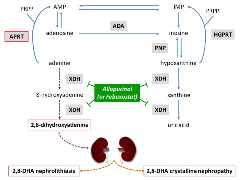
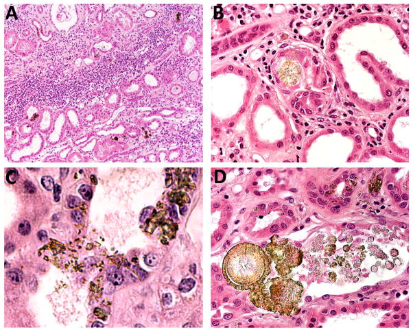
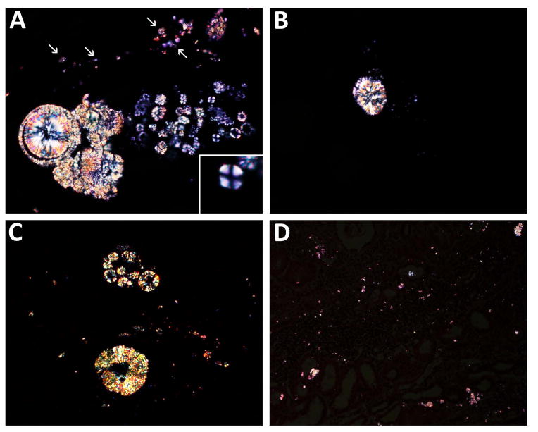
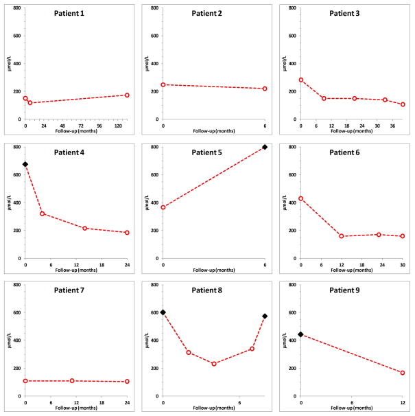
Adenine phosphoribosyltransferase (APRT) deficiency is a rare autosomal recessive enzyme defect of purine metabolism that usually manifests as 2,8-dihydroxyadenine (2,8-DHA) nephrolithiasis and more rarely chronic kidney disease. The disease is most often misdiagnosed and can recur in the renal allograft. We analyzed nine patients with recurrent 2,8-DHA crystalline nephropathy, in all of whom the diagnosis had been missed prior to renal transplantation. The diagnosis was established at a median of 5 (range 1.5-312) weeks following the transplant procedure. Patients had delayed graft function (n=2), acute-on-chronic (n=5) or acute (n=1) allograft dysfunction, whereas one patient had normal graft function at the time of diagnosis. Analysis of allograft biopsies showed birefringent 2,8-DHA crystals in renal tubular lumens, within tubular epithelial cells and interstitium. Fourier transformed infrared microscopy confirmed the diagnosis in all cases, which was further supported by 2,8-DHA crystalluria, undetectable erythrocyte APRT enzyme activity, and genetic testing. With allopurinol therapy, the allograft function improved (n=7), remained stable (n=1) or worsened (n=1). At last follow-up, two patients had experienced allograft loss and five had persistent chronic allograft dysfunction. 2,8-DHA nephropathy is a rare but underdiagnosed and preventable disorder that can recur in the renal allograft and may lead to allograft loss.
Keywords: Clinical research/practice; kidney (allograft) function/dysfunction; kidney disease; kidney transplantation/nephrology.
© Copyright 2014 The American Society of Transplantation and the American Society of Transplant Surgeons.
Conflict of interest statement
The authors of this manuscript have no conflicts of interest to disclose as described by the American Journal of Transplantation.
Figures
References
-
- Kamatani N, Terai C, Kuroshima S, Nishioka K, Mikanagi K. Genetic and clinical studies on 19 families with adenine phosphoribosyltransferase deficiencies. Hum Genet. 1987;75:163–8. - PubMed
-
- Edvardsson V, Palsson R, Olafsson I, Hjaltadottir G, Laxdal T. Clinical features and genotype of adenine phosphoribosyltransferase deficiency in iceland. Am J Kidney Dis. 2001;38:473–80. - PubMed
-
- Harambat J, Bollée G, Daudon M, Ceballos-Picot I, Bensman A, Group AS. Adenine phosphoribosyltransferase deficiency in children. Pediatr Nephrol. 2012;27:571–9. - PubMed
-
- de Jong DJ, Assmann KJ, De Abreu RA, Monnens LA, van Liebergen FJ, Dijkman HB, et al. 2,8-Dihydroxyadenine stone formation in a renal transplant recipient due to adenine phosphoribosyltransferase deficiency. J Urol. 1996;156:1754–5. - PubMed
Publication types
MeSH terms
Substances
Supplementary concepts
Grants and funding
LinkOut - more resources
Full Text Sources
Other Literature Sources
Medical
