

Intrinsically disordered tubulin tails: complex tuners of microtubule functions?
- PMID: 25307498
- PMCID: PMC7060838
- DOI: 10.1016/j.semcdb.2014.09.026
Intrinsically disordered tubulin tails: complex tuners of microtubule functions?
Abstract
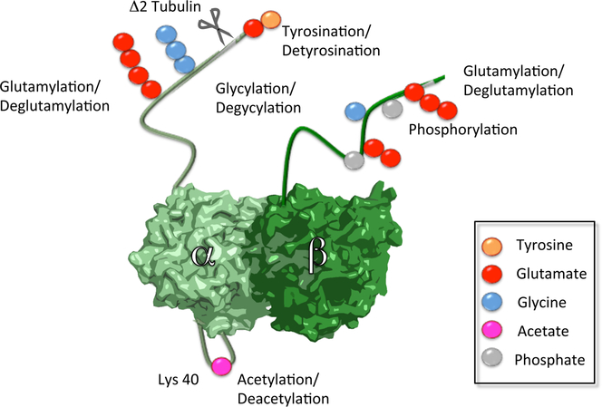
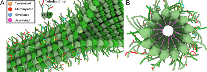
Microtubules are essential cellular polymers assembled from tubulin heterodimers. The tubulin dimer consists of a compact folded globular core and intrinsically disordered C-terminal tails. The tubulin tails form a lawn of densely grafted, negatively charged, flexible peptides on the exterior of the microtubule, potentially akin to brush polymers in the field of synthetic materials. These tails are hotspots for conserved, chemically complex posttranslational modifications that have the potential to act in a combinatorial fashion to regulate microtubule polymer dynamics and interactions with microtubule effectors, giving rise to a "tubulin code". In this review, I summarize our current knowledge of the enzymes that generate the astonishing tubulin chemical diversity observed in cells and describe recent advances in deciphering the roles of tubulin C-terminal tails and their posttranslational modifications in regulating the activity of molecular motors and microtubule associated proteins. Lastly, I outline the promises, challenges and potential pitfalls of deciphering the tubulin code.
Keywords: Brush polymer; Intrinsically disordered proteins; Microtubule; Molecular motors; Post-translational modification; Tubulin tyrosine ligase.
Copyright © 2014. Published by Elsevier Ltd.
Figures
References
-
- Nogales E, Whittaker M, Milligan RA & Downing KH High-resolution model of the microtubule. Cell 96, 79–88 (1999). - PubMed
-
- Mitchison T & Kirschner M Microtubule assembly nucleated by isolated centrosomes. Nature 312, 232–237 (1984). - PubMed
-
- Mitchison T & Kirschner M Dynamic instability of microtubule growth. Nature 312, 237–242 (1984). - PubMed
Publication types
MeSH terms
Substances
Grants and funding
LinkOut - more resources
Full Text Sources
Other Literature Sources
