Review
. 2014:125:477-97.
doi: 10.1016/B978-0-444-62619-6.00027-6.

Current hypotheses on the mechanisms of alcoholism

Affiliations
Review

Current hypotheses on the mechanisms of alcoholism

R P Vetreno et al. Handb Clin Neurol. 2014.

Abstract

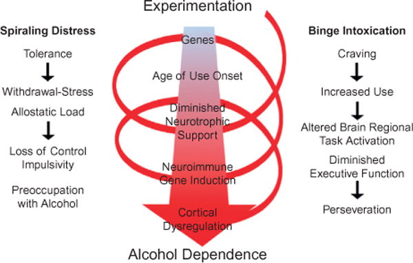
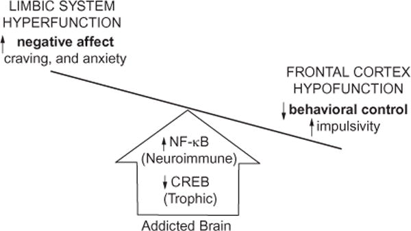
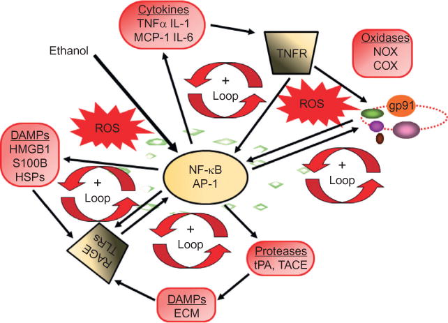
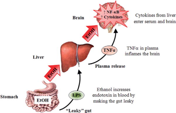
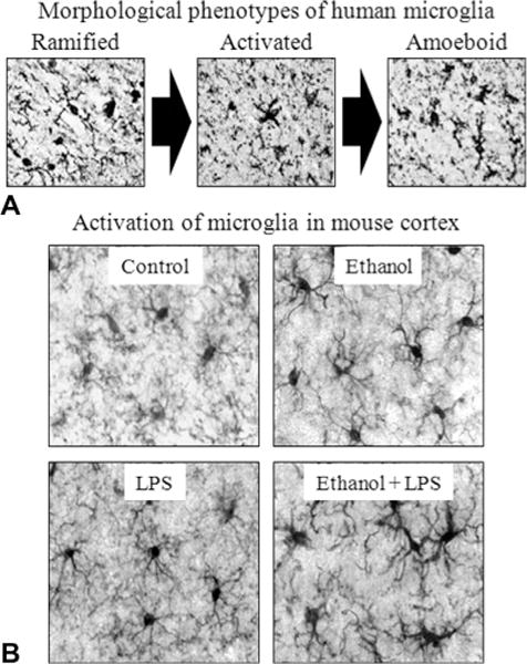
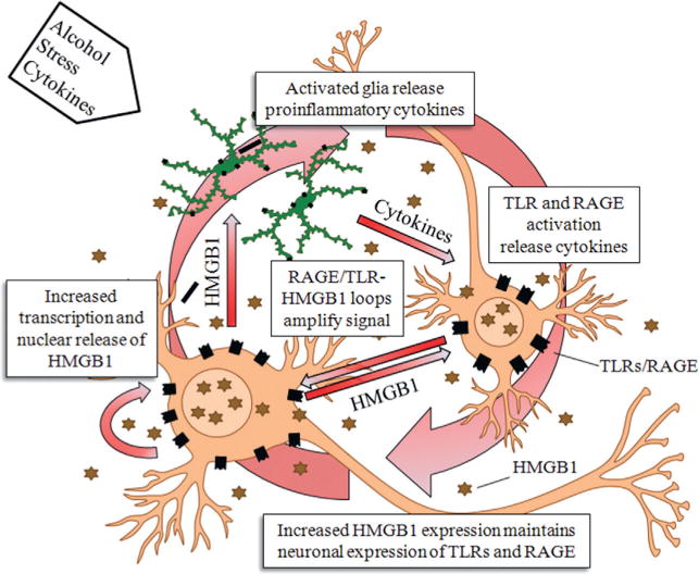
Chronic use of alcohol results in progressive changes to brain and behavior that often lead to the development of alcohol dependence and alcoholism. Although the mechanisms underlying the development of alcoholism remain to be fully elucidated, diminished executive functioning due to hypoactive prefrontal cortex executive control and hyperactive limbic system anxiety and negative emotion might contribute mechanistically to the shift from experimental use to alcoholism and dependence. In the chapter that follows, behavioral deficits associated with cortical dysfunction and neurodegeneration will be related to the behavioral characteristics of alcoholism (e.g., diminished executive function, impulsivity, altered limbic modulation). We will provide evidence that alterations in cyclic AMP-responsive element binding protein (CREB: neurotrophic) and NF-κB (neuroimmune) signaling contribute to the development and persistence of alcoholism. In addition, genetic predispositions and an earlier age of drinking onset will be discussed as contributing factors to the development of alcohol dependence and alcoholism. Overall chronic ethanol-induced neuroimmune gene induction is proposed to alter limbic and frontal neuronal networks contributing to the development and persistence of alcoholism.

Keywords: addiction; alcohol; anxiety; astrocyte; chemokines; cytokines; depression; human; innate immunity; microglia.


Figures

Fig. 27.1
Fig. 27.1
Spiral of distress modeling the progression from alcohol experimentation to alcohol dependence. Left: spiraling distress. Cycles of alcohol intoxication and withdrawal stress increase tolerance and increased allostatic load that promote craving and increased use, leading to further cycles of binge intoxication that progress to loss of control over alcohol use (Koob and Le Moal, 1997; Koob et al., 2004). Middle: Genetics and age of drinking onset are factors that influence binge drinking which, over cycles, alters brain gene expression, e.g., decreased trophic factor expression and increased neuroimmune gene expression that ultimately lead to cortical dysfunction. Right: Craving increases use and binge drinking that disrupts frontal cortical function, reduces behavioral flexibility and executive functions, increasing perseverative-compulsive actions and decreasing goal orientation. The spiral of distress links binge intoxication cycles to alcohol dependence, characterized by preoccupation with alcohol and impulsive drug taking that is likely due in part to diminished frontal cortical executive function. These mechanisms promote alcohol dependence and addiction through increased craving and other negative feelings as well as perseveration and loss of control over drinking. (Adapted from Koob and Le Moal, 1997.)
Fig. 27.2
Fig. 27.2
Comparison of human alcoholic cortical thinning with rat binge alcohol treatment-induced neurodegeneration. Shown are a horizontal section through the ventral rat brain and the ventral surface of the human brain. (Left side) Rat brain anatomic section in the upper left indicates location of ventral horizontal section. The bottom left shows a brain section with silver cell death stain (black areas) showing neurodegeneration from a binge-drinking model that includes association cortical areas, piriform and perirhinal cortex, as well as entorhinal cortex. Blue shading indicates that rat piriform neurodegeneration extends to frontal regions. (Adapted from Obernier et al., 2002a; Crews et al., 2004.) On the right side is a ventral view of the cortex from the human brain. Human alcoholic ventral cortical thinning is shown as a significance map of group differences in cortical thickness in abstinent alcoholics as compared to non-alcoholic controls with areas showing brain regions with significant cortical thinning in alcoholics highlighted in blue (light blue, p<0.01; dark blue, p<0.05). (Adapted from Fortier et al., 2011.) Note perirhinal, entorhinal, and piriform cortex are areas with cortical thinning in humans and areas of binge-drinking neurodegeneration in rats, suggesting binge-drinking human alcoholics are damaging association cortical areas. (Adapted from Fortier et al., 2011.)
Fig. 27.3
Fig. 27.3
Binge drinking reduces behavioral flexibility as assessed by reversal learning. This study treated rats with a 4-day binge-drinking model and then assessed learning and reversal learning after weeks of abstinence to assess long-term changes induced by binge drinking. All animals (both control (CON) and alcohol binge-treated (EtOH)) learned the initial location without any apparent learning differences. Both CON and EtOH animals learned the location of the platform in the Morris water maze task, suggesting binge drinking did not alter learning. For reversal learning the submerged platform was moved to the opposite quadrant and animals were given four trials (21 days postbinge treatment). Binge-treated animals had difficulty. They took more time to locate the new location, spent more time in the previous location consistent with perseveration on the old location, and took longer to reach criteria in the reversal task. (A) Shown is the mean ± SEM of the time required to find the reversal location during the reversal trials. Reversal criterion was set at 2 sd from the distance to platform on the last day of the reference memory task. EtOH animals required a significantly greater number of trials to reach criterion than CON animals [t(14) = 2.376; *p<0.05]. (B) A vertical view of the track taken by a CON and an EtOH rat during the first reversal trial. The open circle represents the location of the submerged platform the animals initially learned. The red circle represents the location of the platform during the reversal learning task. The EtOH animal has numerous reentries into the original goal quadrant due to perseverative behavior and poor search strategy and fails to reach the new platform location within the 90-second time allowed. The binge-treated animals have not received ethanol for several weeks. The reversal learning deficits are consistent with a loss of behavioral flexibility due to disruption of frontal cortical circuits. (Data summarized from Obernier et al., 2002b.)
Fig. 27.4
Fig. 27.4
Alcohol-induced shift in neurotrophic/neuroimmune axis. A simplified schematic distinguishing the frontal cortical and limbic system behavioral changes that characterize drug-induced activation of the innate immune system. Both alcohol addiction and stress upregulate nuclear factor kappa-light-chain enhancer of activated B cells (NF-κB) signaling while reducing cyclic AMP-responsive element-binding protein (CREB) expression, which reduces glutamate transporters, leading to prefrontal cortex hyperexcitability (Crews et al., 2006a; Zou and Crews, 2006; Reissner and Kalivas, 2010) and diminished cortical behavioral control and behavioral inflexibility (Stalnaker et al., 2009; Gruber et al., 2010). Concurrently, induction of innate immune genes in limbic brain regions increases negative affect, craving, and anxiety-like behaviors, prompting further drug abuse and self-medication (Kelley and Dantzer, 2011). The long-term deleterious effects of prolonged alcohol abuse involve diminished activation of frontal cortical behavioral control circuits that lead to a progressive loss of attention and poor decision making that combine with increased negative affect and anxiety that motivate further drug-taking behaviors. Taken together, these innate immune gene-induced behavioral changes characterize the drug-addicted brain (Crews and Vetreno, 2011).
Fig. 27.5
Fig. 27.5
Loops of signaling converge on NF-κB, increasing the expression of chemokines, cytokines, oxidases, and proteases. The proinflammatory transcription factor NF-κB is involved in the induction of neuroimmune genes (Knapp and Crews, 1999; He and Crews, 2008; Qin et al., 2008; Alfonso-Loeches et al., 2010; Zou and Crews, 2010). Alcohol (ethanol) increases NF-κB-DNA binding and transcription. Positive loops of activation occur through induction of genes that stimulate further NF-κB activation, leading to autocrine and paracrine amplification and persistent signals. Cytokines and chemokines, such as TNF-α, IL-1β, IL-6, and MCP-1, as well as their receptors (TNFR in figure), are induced, resulting in amplification loops. Reactive oxygen species (ROS) resulting from oxidases such as NADPH oxidase or ethanol metabolism increase NF-κB transcription of NOX2phox (gp91), a key NOX catalytic subunit (Cao et al., 2005) that produces ROS (Qin et al., 2008). Toll-like receptors (TLRs) and the receptor for advanced glycation end product (RAGE) are increased by ethanol (Alfonso-Loeches et al., 2010; Crews et al., 2013; Vetreno et al., 2013), as are other neuroimmune signaling molecules, resulting in the formation of positive activation loops (Crews et al., 2013; Vetreno et al., 2013). These loops spread neuroimmune signaling across the brain, causing altered neurocircuitry and neurobiology. AP-1, activator protein-1; COX, cyclooxygenase; DAMPs, damage- (or danger-) associated molecular patterns; ECM, extracellular matrix; HMGB1, high-mobility group box 1; HSPs, heat shock proteins; IL-1, interleukin-1; MCP-1, monocyte chemoattractant protein-1; NF-κB, nuclear factor kappa-light-chain-enhancer of activated B cells; NOX, nicotinamide adenine dinucleotide phosphate (NADPH) oxidases; TACE,TNF-α converting enzyme; TNF-α, tissue necrosis factor-alpha; tPA, tissue plasminogen activator.
Fig. 27.6
Fig. 27.6
Systemic ethanol (EtOH) activation of cytokine signaling in the brain. Consumed EtOH enters the stomach and makes it “leaky,” allowing lipopolysaccharide (LPS) to enter the plasma. The circulating EtOH and LPS lead to liver proinflammatory cytokine induction, which results in the production and secretion of tumor necrosis factor-alpha (TNF-α) and other proinflammatory cytokines. TNF-α is transported into the brain from the blood and increases neuroimmune expression through increased activation and the synthesis of nuclear factor kappa-light-chain enhancer of activated B cells (NF-κB) and other cytokines. See Qin et al. (2007) for details.
Fig. 27.7
Fig. 27.7
Microglial morphology, lipopolysaccharide (LPS) and alcohol in the brain. (A) Representative figures depicting characteristic stages of microglial activation. Ramified or “resting” microglia are characterized by long, highly ramified processes with comparatively small cell bodies. Activated microglia are characterized by swollen, truncated processes and enlarged cell bodies. Ameboid or “phagocytic” microglia are characterized by large, ameba-like cell body with no or few small processes (Kreutzberg, 1996; Raivich et al., 1999). Photomicrographs depict stages of microglial activation in postmortem human brain tissue (He and Crews, 2008). (B) Representative figures depicting microglial activation in the mouse cortex following ethanol and/or LPS treatment. Male C57BL/6 mice were treated with saline or ethanol (5 g/kg, i.g.) for 10 days. Half of the subjects received a dose of LPS (3 mg/kg, i.p.) 24 hours after ethanol/saline treatment, and were sacrificed 1 hour later. Ten daily doses of ethanol exposure potentiated LPS-induced microglial activation. (Adapted from Qin et al., 2008.)
Fig. 27.8
Fig. 27.8
Neuroimmune activation through HMGB1-TLR signaling loops. A single injection of lipopolysaccharide (LPS: 5 mg/kg, i.p.) in C57BL/6 mice caused a long-lasting increase in protein expression of tumor necrosis factor-alpha (TNF-α). (See Qin et al., 2007.) The persistent priming of neuroimmune activation lasts for long periods due to positive loops of signaling between neurons and glia. HMGB1 released from neurons stimulates glial activation and glial synthesis of proinflammatory cytokines that further induce additional neuroimmune genes consistent with the long-lasting impact of alcoholism and the persistence, e.g., the folklore that once an alcoholic always an alcoholic (e.g., the neuroimmune priming persists and does not go away, like the risk of relapse to alcoholism). (Adapted from Vetreno et al., 2013.)
Fig. 27.9
Fig. 27.9
Chronic ethanol (EtOH) self-administration induces depression-like behavior and inhibits hippocampal neurogenesis. C57BL/6J mice self-administered either ethanol (10% v/v) or water for 28 days. (A) Increased immobility (seconds) on the forced-swim test provides an index of depression-like behavior. Following a period of abstinence from chronic ethanol consumption, mice evidenced increased immobility time relative to controls. (B) Doublecortin (DCX) expression, a marker of neurogenesis, was reduced in the dentate gyrus of mice exposed to chronic ethanol self-administration. (C) Representative photomicrographs depicting the reduced DCX expression. Alcohol-induced reduced neurogenesis and neuroprogenitor cell proliferation are associated with increased depression-like behavior. Antidepressants reverse both (Stevenson et al., 2009). Scale bar = 200 mm. **p<0.01, relative to control. (Adapted from Stevenson et al., 2009.)
Fig. 27.10
Fig. 27.10
Neuroimmune signaling cascade activation and evidence for involvement in alcohol-induced neurodegeneration. A simplified schematic of the TLR and RAGE receptor signaling cascades. Stimulation of TLRs leads to the generation of ROS and downstream activation of NF-κB. Similarly, activation of the RAGE receptor leads to caspase-3 induction and downstream activation of NF-κB. The production of NF-κB leads to the secretion of proinflammatory gene expression, neuroimmune induction, and cell death. AP-1, activator protein-1; CD14, cluster of differentiation 14; ERK, extracellular signal-regulated kinase; HMGB1, high-mobility group box-1; IKK, inhibitor of nuclear factor kappa-B; JNK, c-Jun N-terminal kinases; LPS, lipopolysaccharide; MyD88, myeloid differentiation primary response gene 88; NADPH oxidase, nicotinamide adenine dinucleotide phosphate-oxidase; NF-κB, nuclear factor kappa-light-chain enhancer of activated B cells; RAGE, receptor for advanced glycation end products; ROS, reactive oxygen species; S100B, S100 calcium-binding protein B; Src, proto-oncogene tyrosine-protein kinase; TIRAP, Toll/interleukin-1 receptor domain-containing adaptor protein; TLRs, Toll-like receptors.
Fig. 27.11
Fig. 27.11
Hyperexcitability contributes to the neurobiology of addiction. A simplified schematic depicting how neuroimmune signaling leads to hyperexcitability and the neurobiology of addiction. Alcohol and stress activate neurons and glia in the central nervous system, resulting in the release of various neuroimmune signals (e.g., high-mobility group box 1 (HMGB1) and interleukin-1β (IL-1β)) that activate neuroimmune receptors (i.e., Toll-like receptors (TLRs) and receptor for advanced glycation end products (RAGE)). Neuroimmune receptor stimulation leads to activation of glutamatergic N-methyl-D-aspartate (NMDA) receptors, e.g., NR2B (Maroso et al., 2010; Iori et al., 2013), which increases Ca2+ flux, triggering induction of neuroimmune genes. In addition, TLR/RAGE activation leads to downstream transcription of nuclear factor kappa-light-chain enhancer of activated B cells (NF-κB) signaling that might be accompanied by diminished cyclic AMP-responsive element-binding protein (CREB) expression, which contributes to neuroimmune gene induction. These two pathways converge, leading to cycles of neuroimmune gene induction that lead to hyperexcitability, neuronal cell death, and network reorganization that culminates in addiction.
Fig. 27.12
Fig. 27.12
Receptor for advanced glycation end product (RAGE), Toll-like receptor (TLR) 3 and 4, and high-mobility group box 1 (HMGB1) expression in the human alcoholic postmortem orbitofrontal cortex correlates with age of drinking onset. The younger the age of drinking onset, the greater the risks of developing alcohol dependence (Grant, 1998). Neuroimmune signaling shows a similar correlation consistent with alcohol-induced increased neuroimmune signaling, increasing the risk for alcoholism. Depicted are individual self and family reports of age of drinking onset vs RAGE (×10 000 pixels/mm2), TLR3 (cells/mm3), TLR4 (cells/mm3), and HMGB1 (cells/mm3) immunoreactivity. Across subjects, age of drinking onset negatively correlated with neuroimmune signal immunoreactivity (r = −0.42, p<0.001). Moderate alcohol drinking controls (CON) tended to self-report a later age of drinking onset (25 ± 1 years of age) in comparison to individuals that met criteria for alcoholism (18 ± 1 years of age). (Adapted from Vetreno et al., 2013).
Fig. 27.13
Fig. 27.13
Receptor for advanced glycation end product (RAGE) and Toll-like receptor 4 (TLR4) expression correlates with high-mobility group box 1 (HMGB1) expression in the prefrontal cortex. The persistent nature of neuroimmune gene induction is likely due to upregulation of both the agonist, HMGB1, and receptors, e.g., TLR receptors that progressively increase in concert with chronic alcohol abuse. (A) Depicted are correlations of individual human RAGE (×10 000 pixels/mm2) and TLR4 (cells/mm3) immunoreactivity versus HMGB1 (cells/mm3) expression in the orbitofrontal cortex (OFC) of alcoholics and moderate-drinking controls. Across subjects, RAGE and TLR4 expression correlated with HMGB1. The correlation of HMGB1 with RAGE/TLR4 is consistent with neuroimmune loops of amplification. (B) Depicted are correlations of RAGE (×10 000 pixels/mm2) and TLR4 (cells/mm2) immunoreactivity versus HMGB1 (cells/mm2) expression in the OFC of adult rats exposed to adolescent binge ethanol. Across subjects, RAGE and TLR4 expression correlated with HMGB1. The correlation of RAGE/TLR4 with HMGB1 indicates the persistence of neuroimmune loops of activation. (Adapted from Vetreno and Crews, 2012; Crews et al., 2013; Vetreno et al., 2013).

References

    1. Akira S, Takeda K, Kaisho T. Toll-like receptors: critical proteins linking innate and acquired immunity. Nat Immunol. 2001;2:675–680. - PubMed
    1. Alfonso-Loeches S, Pascual-Lucas M, Blanco AM, et al. Pivotal role of TLR4 receptors in alcohol-induced neuroinflammation and brain damage. J Neurosci. 2010;30:8285–8295. - PMC - PubMed
    1. Altman J, DAS GD. Autoradiographic and histological evidence of postnatal hippocampal neurogenesis in rats. J Comp Neurol. 1965;124:319–335. - PubMed
    1. APA. Diagnostic and statistical manual of mental disorders: DSM-IV-TR. Washington, D.C: American Psychiatric Association; 2000.
    1. American Psychiatric Association. Diagnostic and statistical manual of mental disorders. 5th. American Psychiatric Publishing; Arlington, VA: 2013.

MeSH terms

Substances