Functional connectivity and neuronal variability of resting state activity in bipolar disorder--reduction and decoupling in anterior cortical midline structures
- PMID: 25307723
- PMCID: PMC6869107
- DOI: 10.1002/hbm.22655
Functional connectivity and neuronal variability of resting state activity in bipolar disorder--reduction and decoupling in anterior cortical midline structures
Abstract
Introduction: The cortical midline structures seem to be involved in the modulation of different resting state networks, such as the default mode network (DMN) and salience network (SN). Alterations in these systems, in particular in the perigenual anterior cingulate cortex (PACC), seem to play a central role in bipolar disorder (BD). However, the exact role of the PACC, and its functional connections to other midline regions (within and outside DMN) still remains unclear in BD.
Methods: We investigated functional connectivity (FC), standard deviation (SD, as a measure of neuronal variability) and their correlation in bipolar patients (n = 40) versus healthy controls (n = 40), in the PACC and in its connections in different frequency bands (standard: 0.01-0.10 Hz; Slow-5: 0.01-0.027 Hz; Slow-4: 0.027-0.073 Hz). Finally, we studied the correlations between FC alterations and clinical-neuropsychological parameters and we explored whether subgroups of patients in different phases of the illness present different patterns of FC abnormalities.
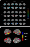
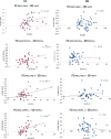
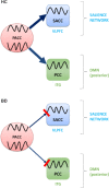
Results: We found in BD decreased FC (especially in Slow-5) from the PACC to other regions located predominantly in the posterior DMN (such as the posterior cingulate cortex (PCC) and inferior temporal gyrus) and in the SN (such as the supragenual anterior cingulate cortex and ventrolateral prefrontal cortex). Second, we found in BD a decoupling between PACC-based FC and variability in the various target regions (without alteration in variability itself). Finally, in our subgroups explorative analysis, we found a decrease in FC between the PACC and supragenual ACC (in depressive phase) and between the PACC and PCC (in manic phase).
Conclusions: These findings suggest that in BD the communication, that is, information transfer, between the different cortical midline regions within the cingulate gyrus does not seem to work properly. This may result in dysbalance between different resting state networks like the DMN and SN. A deficit in the anterior DMN-SN connectivity could lead to an abnormal shifting toward the DMN, while a deficit in the anterior DMN-posterior DMN connectivity could lead to an abnormal shifting toward the SN, resulting in excessive focusing on internal contents and reduced transition from idea to action or in excessive focusing on external contents and increased transition from idea to action, respectively, which could represent central dimensions of depression and mania. If confirmed, they could represent diagnostic markers in BD.
Keywords: bipolar disorder; default mode network; functional connectivity; neuronal variability; perigenual anterior cingulate cortex; resting state fMRI.
© 2014 Wiley Periodicals, Inc.
Figures


References
-
- Akiskal HS (1996): The prevalent clinical spectrum of bipolar disorders: beyond DSM‐IV. J Clin Psychopharmacol 16(Suppl 1):4S–14S. - PubMed
-
- American Psychiatrich Association (1994): Diagnostic and Statistical Manual for Mental Disorders, 4th ed. Washington: American Psychiatrich Association.
-
- Arana GW, Rosenbaum, JF (2000): Handbook of Psychiatric Drug Therapy, 4th ed. Philadelphia: Lippincott Williams & Wilkins.
MeSH terms
LinkOut - more resources
Full Text Sources
Other Literature Sources
Medical

