Review
. 2014 Dec 19:194:76-89.
doi: 10.1016/j.virusres.2014.10.001. Epub 2014 Oct 13.

RNA structure analysis of alphacoronavirus terminal genome regions

Affiliations
Review

RNA structure analysis of alphacoronavirus terminal genome regions

Ramakanth Madhugiri et al. Virus Res. .

Abstract

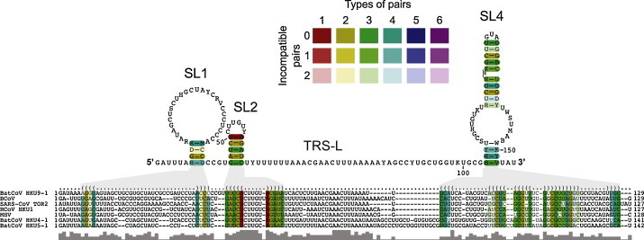
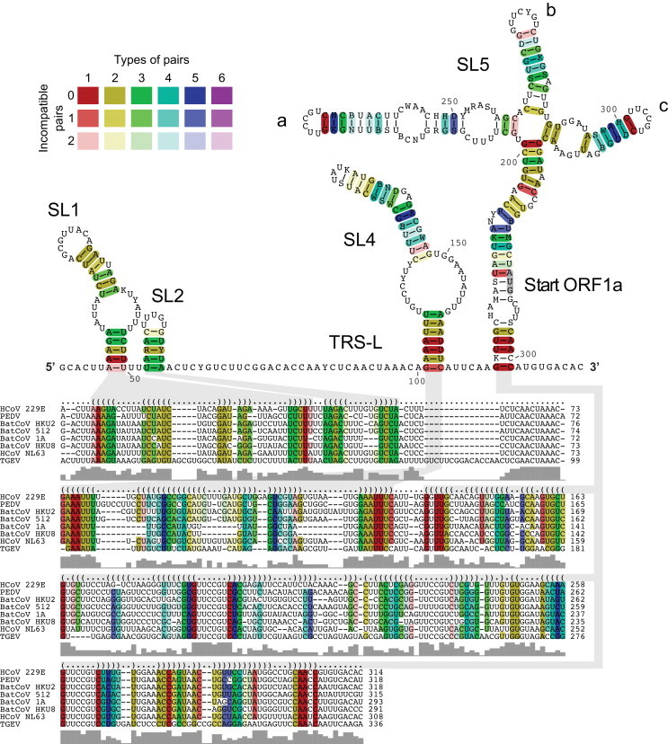
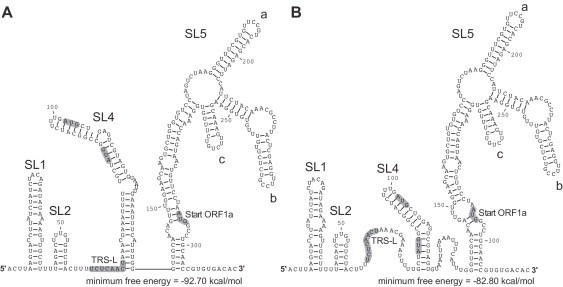
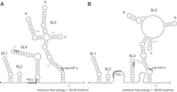
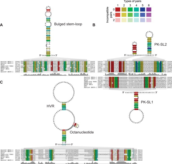
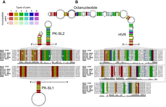
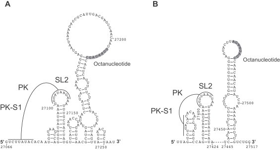
Coronavirus genome replication is mediated by a multi-subunit protein complex that is comprised of more than a dozen virally encoded and several cellular proteins. Interactions of the viral replicase complex with cis-acting RNA elements located in the 5' and 3'-terminal genome regions ensure the specific replication of viral RNA. Over the past years, boundaries and structures of cis-acting RNA elements required for coronavirus genome replication have been extensively characterized in betacoronaviruses and, to a lesser extent, other coronavirus genera. Here, we review our current understanding of coronavirus cis-acting elements located in the terminal genome regions and use a combination of bioinformatic and RNA structure probing studies to identify and characterize putative cis-acting RNA elements in alphacoronaviruses. The study suggests significant RNA structure conservation among members of the genus Alphacoronavirus but also across genus boundaries. Overall, the conservation pattern identified for 5' and 3'-terminal RNA structural elements in the genomes of alpha- and betacoronaviruses is in agreement with the widely used replicase polyprotein-based classification of the Coronavirinae, suggesting co-evolution of the coronavirus replication machinery with cognate cis-acting RNA elements.

Keywords: Coronavirus; RNA structure; RNA virus; Replication; cis-Acting element.


Figures

Fig. 1
Fig. 1
Alignment-based secondary structure prediction of 5′ genome regions of betacoronaviruses. The viruses included in this analysis represent all currently recognized lineages and species in the genus Betacoronavirus. The alignment was generated using LocARNA and the structure was calculated using RNAalifold. The consensus sequence is represented using the IUPAC code: A (adenine), C (cytosine), G (guanine), U (uracil), R (purine [A or G]), Y (pyrimidine [C or U]), M (C or A), K (U or G), W (U or A), S (C or G), B (C, U, or G [not A]), D (A, U, or G [not C]), H (A, U, or C [not G]), V (A, C, or G [not U], N (any base). Colors are used to indicate conserved base pairs: from red (conservation of only one base-pair type) to purple (all six base-pair types are found); from dark (all sequences contain this base pair) to light colors (1 or 2 sequences are unable to form this base pair). The gray bars below the alignment indicate the extent of sequence conservation. Gray shadows are used to link RNA structures with the corresponding dot-bracket notations above the alignment. To refine the alignment, an anchor at the highly conserved apical loop of SL2 was used.
Fig. 2
Fig. 2
Alignment-based secondary structure prediction of 5′ genome regions of alphacoronaviruses. The viruses included in this analysis represent all currently recognized species in the genus Alphacoronavirus. The alignment was calculated by LocARNA, the structure by RNAalifold. The consensus sequence is represented using the IUPAC code. Colors are used to indicate conserved base pairs: from red (conservation of only one base-pair type) to purple (all six base-pair types are found); from dark (all sequences contain this base pair) to light colors (1 or 2 sequences are unable to form this base pair). The gray bars below the alignment indicate the extent of sequence conservation at a given position. Gray shadows are used to link RNA structures with the corresponding dot-bracket notations above the alignment. To refine the alignment, an anchor at the highly conserved core TRS-L was used.
Fig. 3
Fig. 3
RNA secondary structure of the HCoV-229E 5′ UTR. (A) The RNA secondary structure of the 5′ UTR + 20 nts was predicted using RNAfold --noLP. (B) RNA secondary structure of the 5′ UTR + 20 nts was predicted using RNAfold --noLP -C. Structure probing data were used as constraints. The TRS-L core sequence and translational start codons are indicated.
Fig. 4
Fig. 4
RNA secondary structure of the HCoV-NL63 5′ UTR. (A) The RNA secondary structure of the 5′ UTR + 20 nts was predicted using RNAfold --noLP. (B) RNA secondary structure of the 5′ UTR + 20 nts was predicted using RNAfold --noLP -C. Structure probing data were used as constraints. The TRS-L core sequence and translational start codons are indicated.
Fig. 5
Fig. 5
Alignment-based secondary structure prediction of betacoronavirus 3′ genome regions. The viruses included in this analysis represent all currently recognized lineages and species in the genus Betacoronavirus. The alignment was generated using LocARNA and the structure was calculated using RNAalifold. The consensus sequence is represented using the IUPAC code. Colors are used to indicate conserved base pairs: from red (conservation of only one base-pair type) to purple (all six base-pair types are found); from dark (all sequences contain this base pair) to light colors (1 or 2 sequences are unable to form this base pair). Gray bars below the alignment indicate the extent of sequence conservation. Gray shadows are used to link RNA structures with the corresponding dot-bracket notations above the alignment. (A) Alignment-based secondary structure prediction of the bulged stem-loop (BSL) in the 3′ UTR. (B) Alignment-based secondary structure prediction of the pseudoknot (PK) region in the 3′ UTR. Note that PK-SL1 is an alternate structure that requires base-pairing interactions between the loop region of PK-SL2 and the basal part of the BSL shown in (A). Formation of the BSL basal part and PK structure, respectively, are mutually exclusive (see text for details). (C) Alignment-based secondary structure prediction of the hypervariable region (HVR) in the 3′ UTR. To refine the alignment, an anchor at the highly conserved octanucleotide sequence was used.
Fig. 6
Fig. 6
Alignment-based secondary structure prediction of alphacoronavirus 3′-terminal genome regions. The viruses included in this analysis represent all currently recognized species in the genus Alphacoronavirus. The alignment was calculated by LocARNA, the structure by RNAalifold. The consensus sequence is represented using the IUPAC code. Colors are used to indicate conserved base pairs: from red (conservation of only one base-pair type) to purple (all six base-pair types are found); from dark (all sequences contain this base pair) to light colors (1 or 2 sequences are unable to form this base pair). Gray bars below the alignment indicate the extent of sequence conservation at a given position. Gray shadows are used to link RNA structures with the corresponding dot-bracket notations above the alignment. (A) Alignment-based secondary structure prediction of the pseudoknot (PK) region in the 3′ UTR. Formation of the stem of PK-SL1 requires base-pairing interactions with the loop region of SL2. Formation of the PK and the two SL structures shown above the alignment are mutually exclusive (see text for details). (B) Alignment-based secondary structure prediction of the hypervariable region (HVR) in the 3′ UTR. To refine the alignment, an anchor at the highly conserved octanucleotide sequence was used.
Fig. 7
Fig. 7
RNA secondary structure predictions of 3′-terminal genome regions of HCoV-229E (A) and HCoV-NL63 (B). Predictions were generated using RNAfold --noLP -C. As constraints, structure probing data were used. Formation of the predicted pseudoknot (PK) requires base-pairing interactions between the loop region of SL2 and an upstream sequence (and, possibly, structural rearrangements), resulting in the formation of PK stem 1 (PK-S1) as indicated. Also shown is the octanucleotide sequence that is conserved across all genera of the Coronavirinae.

Similar articles

Cited by

References

    1. Almazán F., Galán C., Enjuanes L. The nucleoprotein is required for efficient coronavirus genome replication. J. Virol. 2004;78:12683–12688. - PMC - PubMed
    1. Almazán F., González J.M., Pénzes Z., Izeta A., Calvo E., Plana-Durán J., Enjuanes L. Engineering the largest RNA virus genome as an infectious bacterial artificial chromosome. Proc. Natl. Acad. Sci. U.S.A. 2000;97:5516–5521. - PMC - PubMed
    1. Anand K., Palm G.J., Mesters J.R., Siddell S.G., Ziebuhr J., Hilgenfeld R. Structure of coronavirus main proteinase reveals combination of a chymotrypsin fold with an extra alpha-helical domain. EMBO J. 2002;21:3213–3224. - PMC - PubMed
    1. Anand K., Ziebuhr J., Wadhwani P., Mesters J.R., Hilgenfeld R. Coronavirus main proteinase (3CLpro) structure: basis for design of anti-SARS drugs. Science. 2003;300:1763–1767. - PubMed
    1. Baker S.C., Shieh C.K., Soe L.H., Chang M.F., Vannier D.M., Lai M.M. Identification of a domain required for autoproteolytic cleavage of murine coronavirus gene A polyprotein. J. Virol. 1989;63:3693–3699. - PMC - PubMed

Publication types