. 2015 Feb;36(2):695-706.
doi: 10.1002/hbm.22657. Epub 2014 Oct 12.

Reduced CMRO₂ and cerebrovascular reserve in patients with severe intracranial arterial stenosis: a combined multiparametric qBOLD oxygenation and BOLD fMRI study

Affiliations

Reduced CMRO₂ and cerebrovascular reserve in patients with severe intracranial arterial stenosis: a combined multiparametric qBOLD oxygenation and BOLD fMRI study

Julien Bouvier et al. Hum Brain Mapp. 2015 Feb.

Abstract

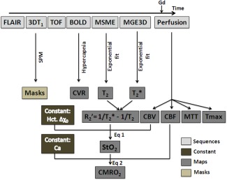
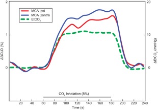
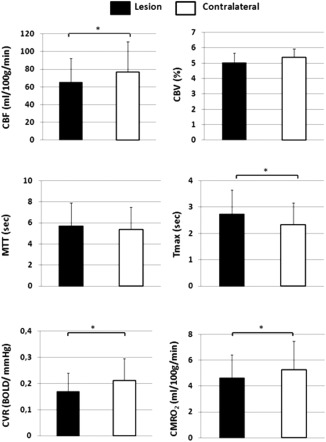
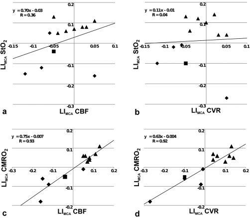
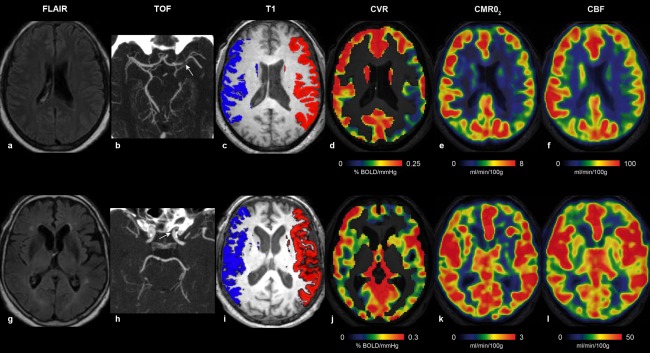
Multiparametric quantitative blood oxygenation level dependent (mqBOLD) magnetic resonance Imaging (MRI) approach allows mapping tissular oxygen saturation (StO2 ) and cerebral metabolic rate of oxygen (CMRO2 ). To identify hemodynamic alteration related to severe intracranial arterial stenosis (SIAS), functional MRI of cerebrovascular reserve (CVR BOLD fMRI) to hypercapnia has been proposed. Diffusion imaging suggests chronic low grade ischemia in patients with impaired CVR. The aim of the present study was to evaluate how oxygen parameters (StO2 and CMRO2 ), assessed with mqBOLD approach, correlate with CVR in patients (n = 12) with SIAS and without arterial occlusion. The perfusion (dynamic susceptibility contrast), oxygenation, and CVR were compared. The MRI protocol conducted at 3T lasted approximately 1 h. Regions of interest measures on maps were delineated on segmented gray matter (GM) of middle cerebral artery territories. We have shown that decreased CVR is spatially associated with decreased CMRO2 in GM of patients with SIAS. Further, the degree of ipsilateral CVR reduction was well-correlated with the amplitude of the CMRO2 deficit. The altered CMRO2 suggests the presence of a moderate ischemia explained by both a decrease in perfusion and in CVR. CVR and mqBOLD method may be helpful in the selection of patients with SIAS to advocate for medical therapy or percutaneous transluminal angioplasty-stenting.

Keywords: BOLD fMRI; brain; cerebral metabolic rate of oxygen (CMRO2); cerebrovascular reserve; hypercapnia; intracranial arterial stenosis; microvasculature; perfusion; stroke; tissular oxygen saturation (StO2).


Figures

Figure 1
Figure 1
Diagram of the acquisition and processing schemes to obtain CVR, perfusion, and oxygenation maps. FLAIR, fluid attenuated inversion recovery; TOF, time of flight; BOLD, blood oxygen level dependent; MSME, multi spin‐echo; MGE3D, 3D multi gradient echo; SPM, statistical parametric mapping; CVR, cerebral vasoreactivity; CBV, cerebral blood volume; CBF, cerebral blood flow; MTT, mean transit time; Tmax, time‐to‐maximum; StO2, tissular oxygen saturation; CMRO2, cerebral metabolic rate of oxygen; Gd, gadolinium‐DOTA; Hct, hematocrit; Δχ 0, change in magnetic susceptibility between oxy and deoxyhemoglobin; Ca, arterial oxygen content. CBV and CBF are obtained from the basal perfusion study. StO2 is derived from R R2 and CBV. CMRO2 is derived from StO2 and CBF. CVR is an independent estimate.
Figure 2
Figure 2
Regressor used for fMRI analyses based on mean EtCO2 variation time courses during hypercapnia (green line). Percentage of BOLD signal averaged over three blocks. Mean BOLD signal time courses of MCA territories ipsilateral and contralateral to the SIAS were represented by red and blue lines, respectively. Both amplitude and initial slope of the BOLD time course decreased in the MCA territory ipsilateral to the SIAS.
Figure 3
Figure 3
(a) CBF, (b) CBV, (c) MTT, (d) Tmax, (e) CVR, and (f) CMRO2 obtained from the SIAS ipsilateral and the contralateral MCA territories and averaged across patients (n = 12) (mean ± SD).
Figure 4
Figure 4
(a) LIMCA_StO2 versus LIMCA_CBF, (b) LIMCA_StO2 versus LIMCA_CVR, (c) LIMCA_CMRO2 versus LIMCA_CBF, and (d) LIMCA_CMRO2 versus LIMCA_CVR (n = 12). StO2, tissular oxygen saturation; CBF, cerebral blood flow; CMRO2, cerebral metabolic rate of oxygen; CVR, cerebrovascular reserve; LIMCA, laterality indices in MCA territories. Each triangle, diamond and square represents patient with right (MCA/ICA), Left (MCA/ICA) or bilateral (MCA/ICA) stenosis, respectively.
Figure 5
Figure 5
Illustrative individual maps of Patient #12 (upper row, a–f) and Patient #4 (lower row, g–l), representing FLAIR (a, g), TOF with stenosis (white arrows) (b, h), ROIs used for delineate the MCA territories overlaid onto axial T 1‐WI (c, i), BOLD response to hypercapnia (d, j), CMRO2 (e, k), and CBF (f, l). Patient #12 had a combined reduction of CBF, CVR, and CMRO2 in the left MCA territory downstream a left MCA SIAS, whereas Patient #4 with a left ICA SIAS had symmetrical values.

References

    1. An H, Lin W (2000): Quantitative measurements of cerebral blood oxygen saturation using magnetic resonance imaging. J Cereb Blood Flow Metab 20:1225–1236. - PMC - PubMed
    1. An H, Lin W, Celik A, Lee YZ (2001): Quantitative measurements of cerebral metabolic rate of oxygen utilization using MRI: A volunteer study. NMR Biomed 14:441–447. - PMC - PubMed
    1. Attye A, Villien M, Tahon F, Warnking J, Detante O, Krainik A (2014): Normalization of cerebral vasoreactivity using BOLD MRI after intravascular stenting. Hum Brain Mapp 35:1320–1324. - PMC - PubMed
    1. Baron JC, Jones T (2012): Oxygen metabolism, oxygen extraction and positron emission tomography: Historical perspective and impact on basic and clinical neuroscience. NeuroImage 61:492–504. - PubMed
    1. Blicher JU, Stagg CJ, O'Shea J, Ostergaard L, MacIntosh BJ, Johansen‐Berg H, Jezzard P, Donahue MJ (2012): Visualization of altered neurovascular coupling in chronic stroke patients using multimodal functional MRI. J Cerebral Blood Flow Metab 32:2044–2054. - PMC - PubMed

Publication types

LinkOut - more resources