

Regression of hepatocellular adenomas with strict dietary therapy in patients with glycogen storage disease type I
- PMID: 25308557
- PMCID: PMC4361930
- DOI: 10.1007/8904_2014_344
Regression of hepatocellular adenomas with strict dietary therapy in patients with glycogen storage disease type I
Abstract
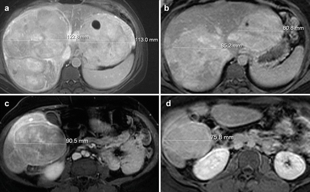
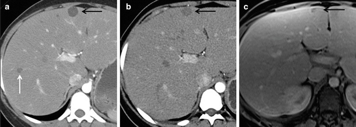
Hepatocellular adenomas (HCAs) are a common complication in patients with glycogen storage disease type I (GSD I). In this series, we report regression of HCAs in a cohort of patients who achieved metabolic control with strict dietary therapy. A retrospective review of the clinical records for all patients with GSD I was performed at our institution. All available imaging studies were reviewed in patients with reported regression of HCAs in the medical record. The charts of 163 patients with GSD Ia and 42 patients with GSD Ib were reviewed, and HCAs were documented in 47 subjects (43 Ia/4 Ib). After review of all available imaging studies, eight patients met criteria of being followed with both magnetic resonance imaging and ultrasound and were found to show evidence of regression of HCAs. In these individuals, regression of the HCAs occurred once metabolic control was obtained, as determined by decreasing levels of serum triglyceride levels. The average triglyceride level in all patients prior to regression of HCAs was 753 mg/dL (SD ± 293). The average serum triglyceride level in all patients at the time of regression of HCAs was 340 mg/dL (SD ± 164). These findings suggest that strict dietary therapy may cause regression of HCAs. If HCAs are documented in a patient with suboptimal metabolic control, intensive medical therapy may be an alternative to surgical intervention in some individuals.
Figures


Similar articles
-
Liver transplantation for glycogen storage disease type Ia.J Hepatol. 2009 Sep;51(3):483-90. doi: 10.1016/j.jhep.2009.05.026. Epub 2009 Jun 17. J Hepatol. 2009. PMID: 19596478
-
Successful treatment of multiple hepatocellular adenomas with percutaneous radiofrequency ablation.World J Gastroenterol. 2013 Nov 14;19(42):7480-6. doi: 10.3748/wjg.v19.i42.7480. World J Gastroenterol. 2013. PMID: 24259982 Free PMC article.
-
Development of hepatocellular adenomas in a patient with glycogen storage disease Ia treated with growth hormone therapy.JIMD Rep. 2023 Aug 18;64(5):303-311. doi: 10.1002/jmd2.12381. eCollection 2023 Sep. JIMD Rep. 2023. PMID: 37701330 Free PMC article.
-
Glycogen storage disease in adults.Ann Intern Med. 1994 Feb 1;120(3):218-26. doi: 10.7326/0003-4819-120-3-199402010-00008. Ann Intern Med. 1994. PMID: 8273986 Review.
-
Paediatric hepatocellular adenomas: Lessons from a systematic review of relevant literature.JHEP Rep. 2024 Mar 27;6(6):101078. doi: 10.1016/j.jhepr.2024.101078. eCollection 2024 Jun. JHEP Rep. 2024. PMID: 38699071 Free PMC article. Review.
Cited by
-
Glycogen storage disease type Ia: Adult presentation with microcytic anemia and liver adenomas.Hepatology. 2018 Aug;68(2):780-782. doi: 10.1002/hep.29858. Epub 2018 Apr 27. Hepatology. 2018. PMID: 29486517 Free PMC article. No abstract available.
-
Elevated Plasma Vitamin B12 in Patients with Hepatic Glycogen Storage Diseases.J Clin Med. 2020 Jul 22;9(8):2326. doi: 10.3390/jcm9082326. J Clin Med. 2020. PMID: 32707782 Free PMC article.
-
Over 20-Year Follow-up of Patients with Hepatic Glycogen Storage Diseases: Single-Center Experience.Diagnostics (Basel). 2020 May 13;10(5):297. doi: 10.3390/diagnostics10050297. Diagnostics (Basel). 2020. PMID: 32414085 Free PMC article.
-
Cell Modeling and Rescue of a Novel Non-coding Genetic Cause of Glycogen Storage Disease IX.bioRxiv [Preprint]. 2025 May 17:2025.05.14.654043. doi: 10.1101/2025.05.14.654043. bioRxiv. 2025. PMID: 40462889 Free PMC article. Preprint.
-
Impaired Very-Low-Density Lipoprotein catabolism links hypoglycemia to hypertriglyceridemia in Glycogen Storage Disease type Ia.J Inherit Metab Dis. 2021 Jul;44(4):879-892. doi: 10.1002/jimd.12380. Epub 2021 Apr 7. J Inherit Metab Dis. 2021. PMID: 33739445 Free PMC article.
References
-
- Ault GT, Wren SM, Ralls PW, Reynolds TB, Stain SC. Selective management of hepatic adenomas. Am Surg. 1996;62:825–829. - PubMed
-
- Bianchi L. Glycogen storage disease I and hepatocellular tumours. Eur J Pediatr. 1993;152(Suppl 1):534–536. - PubMed
-
- Coire CI, Qizilbash AH, Castelli MF. Hepatic adenomata in type Ia glycogen storage disease. Arch Pathol Lab Med. 1987;111:166–169. - PubMed
Grants and funding
LinkOut - more resources
Full Text Sources
Other Literature Sources
Miscellaneous
