. 2015 Apr;17(4):504-19.
doi: 10.1111/cmi.12380. Epub 2014 Nov 22.

The Anaplasma phagocytophilum effector AmpA hijacks host cell SUMOylation

Affiliations

The Anaplasma phagocytophilum effector AmpA hijacks host cell SUMOylation

Andrea R Beyer et al. Cell Microbiol. 2015 Apr.

Abstract

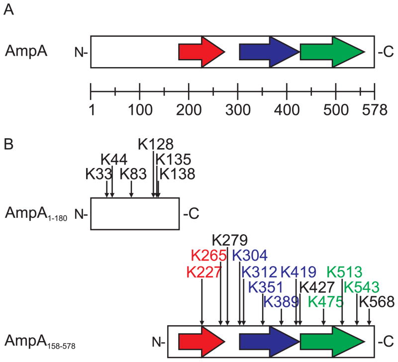
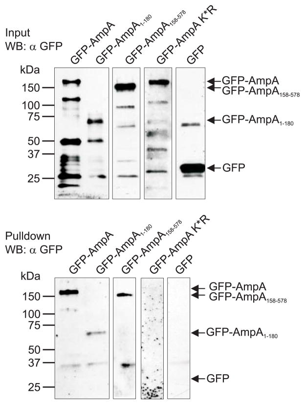
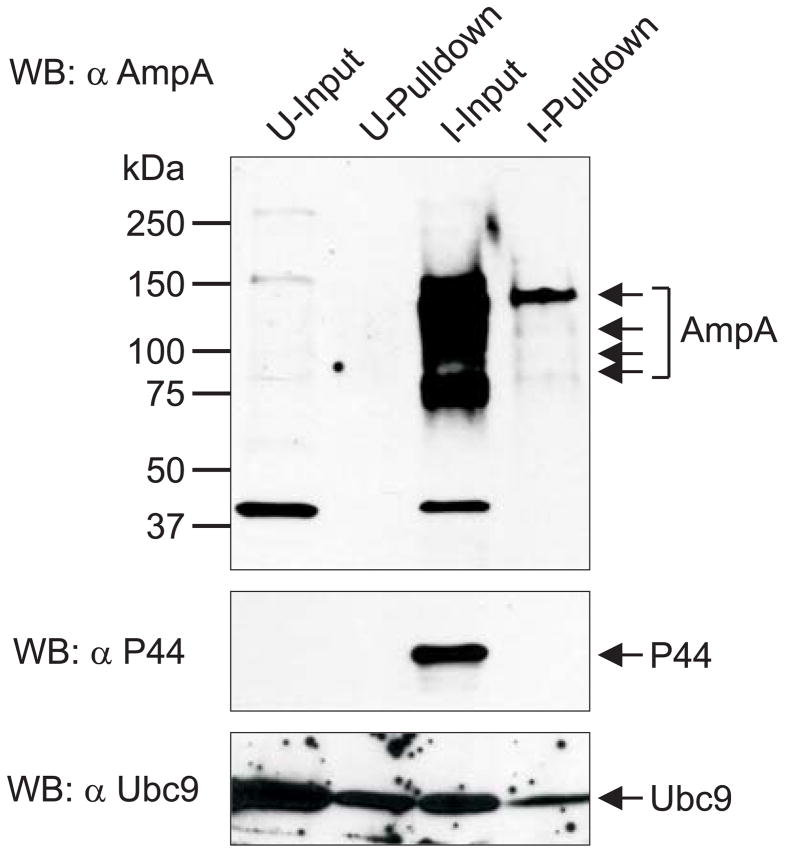
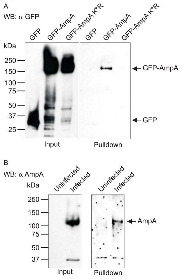
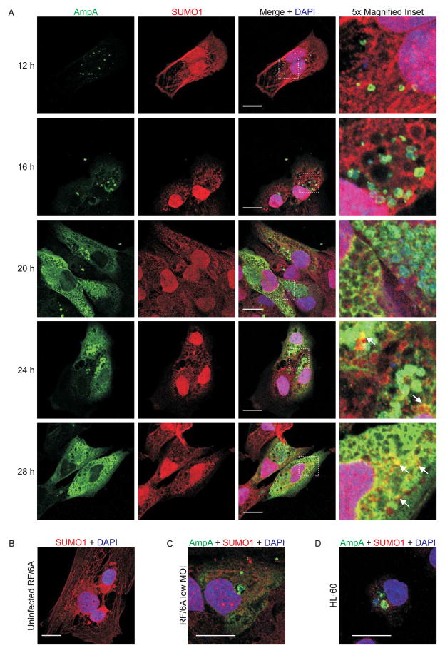
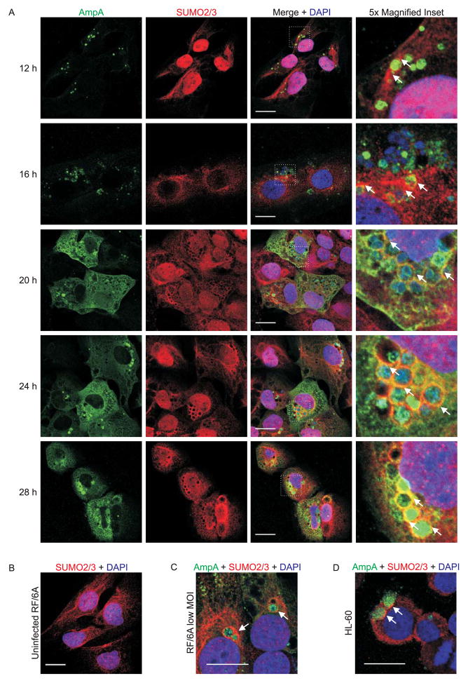
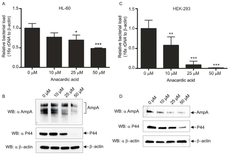
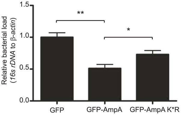
SUMOylation, the covalent attachment of a member of the small ubiquitin-like modifier (SUMO) family of proteins to lysines in target substrates, is an essential post-translational modification in eukaryotes. Microbial manipulation of SUMOylation recently emerged as a key virulence strategy for viruses and facultative intracellular bacteria, the latter of which have only been shown to deploy effectors that negatively regulate SUMOylation. Here, we demonstrate that the obligate intracellular bacterium, Anaplasma phagocytophilum, utilizes an effector, AmpA (A. phagocytophilum post-translationally modified protein A) that becomes SUMOylated in host cells and this is important for the pathogen's survival. We previously discovered that AmpA (formerly APH1387) localizes to the A. phagocytophilum-occupied vacuolar membrane (AVM). Algorithmic prediction analyses denoted AmpA as a candidate for SUMOylation. We verified this phenomenon using a SUMO affinity matrix to precipitate both native AmpA and ectopically expressed green fluorescent protein (GFP)-tagged AmpA. SUMOylation of AmpA was lysine dependent, as SUMO affinity beads failed to precipitate a GFP-AmpA protein when its lysine residues were substituted with arginine. Ectopically expressed and endogenous AmpA were poly-SUMOylated, which was consistent with the observation that AmpA colocalizes with SUMO2/3 at the AVM. Only late during the infection cycle did AmpA colocalize with SUMO1, which terminally caps poly-SUMO2/3 chains. AmpA was also detected in the cytosol of infected host cells, further supporting its secretion and likely participation in interactions that aid pathogen survival. Indeed, whereas siRNA-mediated knockdown of Ubc9 - a necessary enzyme for SUMOylation - slightly bolstered A. phagocytophilum infection, pharmacologically inhibiting SUMOylation in infected cells significantly reduced the bacterial load. Ectopically expressed GFP-AmpA served as a competitive agonist against native AmpA in infected cells, while lysine-deficient GFP-AmpA was less effective, implying that modification of AmpA lysines is important for infection. Collectively, these data show that AmpA becomes directly SUMOylated during infection, representing a novel tactic for A. phagocytophilum survival.


Figures

Fig. 1
Fig. 1
Schematic diagrams of full-length A. phagocytophilum AmpA and truncated recombinant AmpA proteins used in these studies. A. Diagram of full-length AmpA. AmpA comprises an amino (N)-terminal region (amino acids 1 to 179), a tandem repeat region (amino acids 180 to 549), and a short carboxy (C)-terminal region (amino acids 550 to 578). The repeat region consists of 3 tandemly-arranged direct repeats (indicated by the colored arrows) consisting of 93 (red), 122 (blue), and 130 amino acids (green). The scale indicates 50 amino acid intervals. B. Diagrams of AmpA truncations. The AmpA C-terminal truncation AmpA1-180 (top) lacks the tandem repeat region and contains 6 lysine (K) residues within its 180 amino acid length. The AmpA N-terminal truncation AmpA158-578 (bottom) has the non-repeat region removed and contains 13 lysines within its 421 amino acid length. Each AmpA truncation was fused to GFP (not shown) on its N-terminus. Arrows above the diagram denote lysine (K) residues and their amino acid position within AmpA. Colored lysine positions coordinate to their locations within their respective tandem repeat regions (red, blue, and green arrows).
Fig. 2
Fig. 2
Ectopically expressed AmpA is SUMOylated. SUMOylation pulldowns with ectopically expressed GFP-AmpA, GFP-AmpA1-180, GFP-Amp158-578, GFP-AmpA K*R, and GFP in HeLa cells. Western blots of protein input (top) and pulldown eluates (bottom) were screened with GFP antibody. Arrows denote the expected apparent molecular weights for each GFP-AmpA protein and the expected size for GFP. Blots are representative of 2–4 pulldowns performed for each GFP-AmpA protein.
Fig. 3
Fig. 3
Native AmpA is SUMOylated in A. phagocytophilum infected HL-60 cells. Upper panel shows uninfected (U) and infected (I) HL-60 lysate input and pulldown lanes probed by Western blot analysis with AmpA antibody. Middle and lower panels show Western blotting of samples with antibodies against A. phagocytophilum P44 and Ubc9 as negative and positive controls for SUMOylation, respectively. Arrows denote the multiple isoforms of native AmpA and the expected sizes for P44 and Ubc9. Data presented are representative of four experiments with similar results.
Fig. 4
Fig. 4
Ectopically expressed and native AmpA are poly-SUMOylated. A. Input (left panel) and pulldowns (right panel) of HEK-293 cells transfected to express GFP-AmpA, GFP-AmpA K*R, or GFP alone. Western blots were probed with GFP antibody. Arrows denote the expected apparent molecular weights for GFP-AmpA, GFP-AmpA K*R, and GFP. B. Input (left panel) and pulldowns (right panel) of uninfected and A. phagocytophilum infected HL-60 cells. Western blots were probed with AmpA antibody. Arrow denotes the expected apparent molecular weight for native AmpA protein. Data shown are representative of two experiments with similar results.
Fig. 5
Fig. 5
Localization of SUMO1 and AmpA in A. phagocytophilum infected mammalian host cells. A. Representative images of infected RF/6A cells at 12, 16, 20, 24, and 28 h post infection. Time points are marked at the left of each row. Cells were immunofluorescently stained to detect AmpA (green), SUMO1 (red), and DNA (DAPI, blue). Merged images are shown in the third column. Areas demarcated by white hatched boxes in the merged column are magnified five-fold in the fourth column. White arrows indicate representative areas where AmpA and SUMO1 signals colocalize. Scale bars, 20 μm. B. SUMO1 (red) and DNA (DAPI, blue) staining in an uninfected RF/6A cell. C and D. Merged confocal images of RF/6A cells infected with a low MOI of A. phagocytophilum (C) and A. phagocytophilum infected HL-60 cells (D) stained for AmpA (green), SUMO1 (red), and DNA (DAPI, blue) at 24 h post infection.
Fig. 6
Fig. 6
Localization of SUMO2/3 and AmpA in A. phagocytophilum infected mammalian host cells. A. Representative images of infected RF/6A cells at 12, 16, 20, 24, and 28 h post infection. Time points are marked at the left of each row. Cells were immunofluorescently stained to detect AmpA (green), SUMO2/3 (red), and DNA (DAPI, blue). Merged images are shown in the third column. Areas demarcated by white hatched boxes in the merged column are magnified five-fold in the fourth column. B. SUMO2/3 (red) and DNA (DAPI, blue) staining in an uninfected RF/6A cell. C and D. Merged confocal images of RF/6A cells infected with a low MOI of A. phagocytophilum (C) and A. phagocytophilum infected HL-60 cells (D) stained for AmpA (green), SUMO2/3 (red), and DNA (DAPI, blue) at 24 h post infection. White arrows indicate representative areas where AmpA and SUMO2/3 signals colocalize and/or SUMO2/3 accumulates on the AVM. Scale bars, 20 μm.
Fig. 7
Fig. 7
Pharmacologic inhibition of SUMOylation reduces the A. phagocytophilum load. HL-60 (A and B) and HEK-293 (C and D) cells were treated with 0, 10, 25, or 50 μM anacardic acid for 4 h prior to infection by A. phagocytophilum. The infected cells were maintained for 96 h, with anacardic acid-containing growth media being replenished every 24 h. Cells were collected for qPCR (A and C) and Western blot analyses (B and D). For qPCR, relative DNA loads of A. phagocytophilum 16s rRNA gene were normalized to DNA loads of the human β-actin gene using the 2−ΔΔCT (Livak) method. Results shown are the means ± SD of triplicate samples and are representative of three independent experiments in HL-60 cells and two independent experiments in HEK-293 cells, with similar results. Statistically significant (*P < 0.05; **P < 0.005; ***P < 0.001) values relative to the 0 μM control are indicated. In B and D, 15 μg or 5 μg of whole cell lysate, respectively, was loaded per lane. Blots were probed with AmpA and P44 antibodies to examine pathogen burden, in comparison to the β-actin loading control. Data shown are representative of two independent sets of Western assays performed for each cell type.
Fig. 8
Fig. 8
Ubc9 expression is not altered by A. phagocytophilum infection, but knockdown of Ubc9 results in a significantly increased bacterial load. A and B. A. phagocytophilum infection does not alter Ubc9 expression. A. Western-blotted whole cell lysates of uninfected and A. phagocytophilum infected cells were screened with β-actin and Ubc9 antibodies. B. Three pairs of infected and uninfected HL-60 lysates were examined in duplicate by Western blotting and the ratio of Ubc9 to β-actin analyzed by densitometry. “ns”, not significant. C. HEK-293 cells support A. phagocytophilum infection. HEK-293 cells that had been incubated with A. phagocytophilum were stained with DAPI (blue) to denote host cell nuclei (N) and bacteria (*). Cells were also stained with anti-AmpA (red) prior to visualization using LSCM. Scale bar, 10 μm. D–G. Effect of ubc9 knockdown on the A. phagocytophilum load in infected HEK-293 cells. HEK-293 cells were transfected with siRNA targeting ubc9 or gapdh, non-targeting siRNA, or transfection agent only (mock). At 72 h post transfection, the cells were boosted with a second siRNA transfection and infected with A. phagocytophilum. At 48 h post infection, the cells were resuspended and seeded on coverslips for immunofluorescent staining or collected for whole cell lysis and Western blotting analysis. D. Western blots demonstrating siRNA knockdown of Ubc9. Whole cell lysates from mock-, non-targeting siRNA-, gapdh siRNA-, and ubc9 siRNA-transfected cells were screened with antibodies targeting Ubc9 and GAPDH to confirm knockdown, and β-actin as a loading control. Blots are representative of more than four independent siRNA knockdown assays. E–G. The A. phagocytophilum load is modestly higher in host cells in which ubc9 has been knocked down. siRNA-treated and control host cells that had been infected with A. phagocytophilum were collected and processed for qPCR (E) or immunofluorescence microscopy (F and G). E. Relative loads of A. phagocytophilum 16S rRNA gene were normalized to human β-actin using the 2−ΔΔCT (Livak) method. Results shown are the means ± SD of triplicate samples and are representative of two independent experiments with similar results. Statistically significant (*P < 0.05) values relative to the ubc9 knockdown are indicated. F and G. Coverslips containing siRNA knockdown cells were processed for immunofluorescence and screened with anti-P44 to denote ApVs inside host cells. The mean ± standard deviations of percentages of infected HL-60 cells (F) and Ap vacuolar inclusions per HL-60 cell (G) were determined using immunofluorescence microscopy. Data presented are representative of two independent experiments. Statistically significant (*P < 0.05; **P < 0.005) values are indicated.
Fig. 9
Fig. 9
Ectopically expressed AmpA limits A. phagocytophilum growth in a lysine-dependent manner. Transfected HEK-293 cells expressing GFP-AmpA, GFP-Amp K*R, or GFP were infected with A. phagocytophilum. Genomic DNA was isolated and subjected to qPCR. Relative DNA loads of the A. phagocytophilum 16s rRNA gene were normalized to DNA loads of the human β-actin gene using the 2−ΔΔCT (Livak) method. Results shown are the means ± SD of triplicate samples and are representative of two independent experiments with similar results. Statistically significant (*P < 0.05; **P < 0.005) values relative to the GFP or GFP-AmpA are indicated. GFP vector alone was set as the control, equal to 1.0.

References

    1. Bekes M, Drag M. Trojan horse strategies used by pathogens to influence the small ubiquitin-like modifier (SUMO) system of host eukaryotic cells. J Innate Immun. 2012;4:159–167. - PMC - PubMed
    1. Carlyon JA. Establishing intracellular infection: modulation of host cell functions (Anaplasmataceae) In: Palmer GH, Azad A, editors. Intracellular Pathogens II: Rickettsiales. Washington, D. C: ASM Press; 2012.
    1. Carlyon JA, Chan WT, Galan J, Roos D, Fikrig E. Repression of rac2 mRNA expression by Anaplasma phagocytophila is essential to the inhibition of superoxide production and bacterial proliferation. J Immunol. 2002;169:7009–7018. - PubMed
    1. Cortes C, Rzomp KA, Tvinnereim A, Scidmore MA, Wizel B. Chlamydia pneumoniae inclusion membrane protein Cpn0585 interacts with multiple Rab GTPases. Infect Immun. 2007;75:5586–5596. - PMC - PubMed
    1. Dunphy PS, Luo T, McBride JW. Ehrlichia chaffeensis Exploits Host SUMOylation Pathways To Mediate Effector-Host Interactions and Promote Intracellular Survival. Infect Immun. 2014;82:4154–4168. - PMC - PubMed

Publication types

Substances