Review
. 2015;11(2):149-56.
doi: 10.2174/1573403x10666141013122400.

Atrial tachycardias following atrial fibrillation ablation

Affiliations
Review

Atrial tachycardias following atrial fibrillation ablation

László Sághy et al. Curr Cardiol Rev. 2015.

Abstract

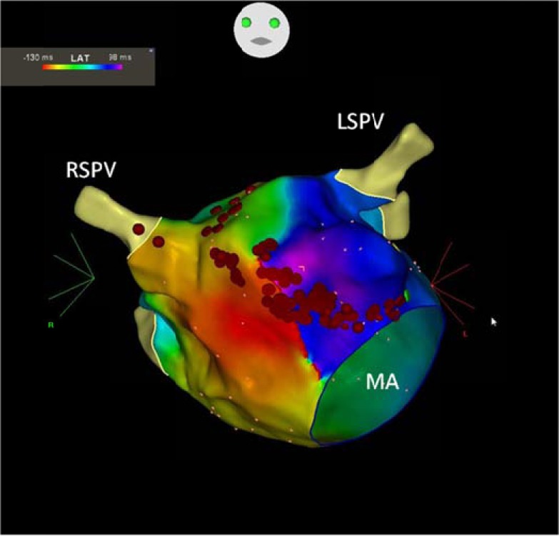
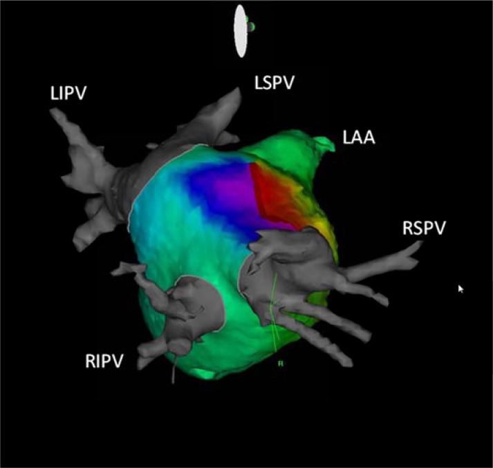
One of the most important proarrhythmic complications after left atrial (LA) ablation is regular atrial tachycardia (AT) or flutter. Those tachycardias that occur after atrial fibrillation (AF) ablation can cause even more severe symptoms than those from the original arrhythmia prior to the index ablation procedure since they are often incessant and associated with rapid ventricular response. Depending on the method and extent of LA ablation and on the electrophysiological properties of underlying LA substrate, the reported incidence of late ATs is variable. To establish the exact mechanism of these tachycardias can be difficult and controversial but correlates with the ablation technique and in the vast majority of cases the mechanism is reentry related to gaps in prior ablation lines. When tachycardias occur, conservative therapy usually is not effective, radiofrequency ablation procedure is mostly successful, but can be challenging, and requires a complex approach.


Figures

Fig. (1)
Fig. (1)
Intracardiac echocardiography image of left atrium. Ablation catheter (ABL) is sitting on the carina between left superior (LSPV) and left inferior (LIPV) pulmonary veins. Lasso: circular catheter in LSPV.
Fig. (2)
Fig. (2)
Twelve lead surface ECG of a narrow isthmus reentry, originating from the vicinity of right inferior pulmonary vein. Note the long isoelectric interval between discrete flutter waves.
Fig. (3)
Fig. (3)
Rapid, regular pulmonary vein tachycardia persists within an isolated right superior pulmonary vein. The atria, outside the isolated vein are in sinus rhythm. (A). Radiofrequency ablation at the earliest spot within the vein, indicated by the ablation artifact on the 4-5 bipoles of Lasso catheter results in the termination of tachycardia. (B) Ls: lasso catheter, CS: coronary sinus catheter, Map d: mapping catheter.
Fig. (4)
Fig. (4)
Gap related tachycardia originating from the circular lesion around the right inferior pulmonary vein. (A, 200mm/sec), and the corresponding catheter positions, arrows point out the direction of activation through two gaps (at lasso pole number 4-5 and 7-8) in the ablation line (B). Entrainment pacing with 245 msec from the earliest lasso bipole (4-5) showed concealed fusion and PPI-TCL was 11 msec. (C). Termination of tachycardia during ablation at the entrance (pole 4-5) (D,100 mm/sec). The vein was isolated with the second ablation at the pole 7-8 (not shown). RIPV: right inferior pulmonary vein, CT: crista terminalis catheter, ICE: intracardiac echocardiography probe, Abl: ablation catheter, Lasso: lasso catheter, CS: coronary sinus catheter, PPI: post pacing interval, TCL: tachycardia cycle length.
Fig. (5)
Fig. (5)
Perimitral reentry propagating around the mitral valve in counterclokwise direction on three dimensional CARTO activation map. Red dots represent the ablation line which was created between the anterior mitral annulus and the right superior pulmonary vein. MA: mitral annulus, RSPV: right superior pulmonary vein, LSPV: left superior pulmonary vein.
Fig. (6)
Fig. (6)
Roof dependent flutter propagating around the right sided pulmonary veins on three dimensional CARTO activation map with CT integration in right lateral view. RSPV: right superior pulmonary vein, RIPV: right inferior pulmonary vein, LSPV: left superior pulmonary vein, LIPV: left inferior pulmonary vein, LAA: left atrial appendage.

References

    1. Oral H, Knight BP, Tada H , et al. Pulmonary vein isolation for paroxysmal and persistent atrial fibrillation. Circulation. 2002;105:1077–81. - PubMed
    1. Oral H, Knight BP, Morady F. Left atrial flutter after segmental ostial radiofrequency catheter ablation for pulmonary vein isolation. Pacing Clin Electrophysiol. 2003;26:1417–9. - PubMed
    1. Gerstenfeld EP, Callans DJ, Dixit S , et al. Mechanisms of organized left atrial tachycardias occurring after pulmonary vein isolation. Circulation. 2004;110:1351–7. - PubMed
    1. Oral H, Scharf C, Chugh A , et al. Catheter ablation for paroxysmal atrial fibrillation Segmental pulmonary vein ostial ablation versus left atrial ablation. Circulation. 2003;108:2355–60. - PubMed
    1. Gaita F, Caponi D, Scaglione M , et al. Long-term clinical results of 2 different ablation strategies in patients with paroxysmal and persistent atrial fibrillation. Circ Arrhythmia Electrophysiol. 2008;1:269–75. - PubMed