. 2014 Sep 12;58(3):2403.
doi: 10.4081/ejh.2014.2403.

Assessment of metal contaminants in non-small cell lung cancer by EDX microanalysis

Affiliations

Assessment of metal contaminants in non-small cell lung cancer by EDX microanalysis

M Scimeca et al. Eur J Histochem. .

Abstract

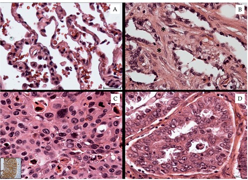
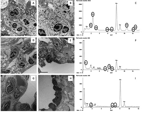
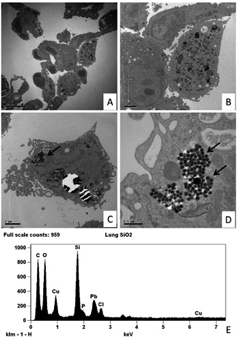
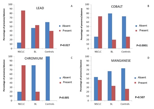
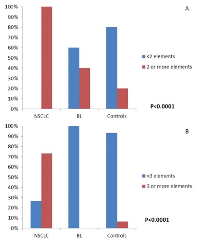
Human cardio-respiratory diseases are strongly correlated to concentrations of atmospheric elements. Bioaccumulation of heavy metals is strictly monitored, because of its possible toxic effects. In this work, we utilized the EDX microanalysis in order to identify the potential heavy metal accumulation in the lung tissue. To this aim, we enrolled 45 human lung biopsies: 15 non-small cell lung cancers, 15 lung benign lesions and 15 control biopsies. Lung samples were both paraffin embedded for light microscopy study and eponepoxid embedded for transmission electron microscopy. EDX microanalysis was performed on 100 nm thick unstained ultrathin-sections placed on specific copper grids. Our results demonstrated that the EDX technology was particularly efficient in the study of elemental composition of lung tissues, where we found heavy metals, such as Cobalt (Co), Chromium (Cr), Manganese (Mn) and Lead (Pb). Furthermore, in malignant lesions we demonstrated the presence of multiple bio-accumulated elements. In fact, a high rate of lung cancers was associated with the presence of 3 or more bio-accumulated elements compared to benign lesions and control tissue (91.7%, 0%, 8.3%, respectively). The environmental impact on pulmonary carcinogenesis could be better clarified by demonstrating the presence of polluting agents in lung tissues. The application of EDX microanalysis on biological tissuescould shed new light in the study of the possible bioaccumulation of polluting agents in different human organs and systems.


Conflict of interest statement

Conflict of interests: the authors declare no conflict of interest.

Figures

Figure 1.
Figure 1.
Lung tissue morphological study. A) Normal lung tissue characterized by alveoli with thin walls. B) Pulmonary fibrosis; asterisks indicate collagen deposition in the alveolar septa. C) Squamous cell carcinoma (inset shows Cytokeratin 5/6 positivity). D) Well differentiated adenocarcinoma.
Figure 2.
Figure 2.
Ultrastructural and EDX analysis of lung tissues. A,B) Lung adenocarcinoma cells with dense granules 400 to 600 nm in diameter (arrow) and large nucleoli. C) Toxic elements detected in one case of NSCLC. D) Inflammatory infiltrates (asterisk indicate a mastocyte) in interstitium of BL tissue. E) Chronic interstitial pneumonia; note lung cells and collagen deposition (asterisk). F) Toxic elements detected in one case of benign lesion. G,H) Lung tissues without significant pulmonary pathology. I) Toxic elements detected in one case of control tissues.
Figure 3.
Figure 3.
Transmission electron microscopy and EDX microanalysis of murine lung tissues. A) Low magnification of thin walled alveoli (15 day of SiO2 exposure). B) Histiocyte with a moderate amount of electron dense granules. C,D) Mice lung electron micrographs after 50 day of SiO2 exposure show an elevated amount of small particles (arrows) into the cytoplasm of histiocytes. E) EDX spectrum shows that the particles indicated in the panel D are made of Si. Other elements can be referred to the composition of the support grid (Cu), staining solution (Pb) and biological tissue (Cl, C, O and P).
Figure 4.
Figure 4.
Elemental analysis. Percentage distributions of the four heavy metal according to the three group of tissues: A) lead (Pb); B): cobalt (Co); C): chromium (Cr); D): manganese (Mn). The presence (red column) and absence (blu column) of heavy metal revealed respectively in Non Small Cell Lung Cancer (NSCLC), benign lesions (BL) and control cases (Controls) are reported as percentage. *Chi-Square’s test. P-values refer to comparisons among the three groups of tissues according to the presence of each specific element.
Figure 5.
Figure 5.
Percentage distribution of the number of heavy metals according to the three groups of tissues: Non Small Cell Lung Cancer (NSCLC), benign lesions (BL) and control cases (Controls). *Chi-Square’s test. P-values refer to comparisons among the three groups of tissues according to the simultaneous presence of elements: 2 or more elements, red column; less than 2, blue columns.

References

    1. Raaschou-Nielsen O, Andersen ZJ, Beelen R, Samoli E, Stafoggia M, Weinmayr G, et al. Air pollution and lung cancer incidence in 17 European cohorts: prospective analyses from the European Study of Cohorts for Air Pollution Effects (ESCAPE). Lancet Oncol 2013;14:813-22 - PubMed
    1. Schottenfeld D, Fraumeni JF., Jr.Cancer epidemiology and prevention, 3rd ed. Oxford, Oxford University Press: 2006
    1. Pope CA, 3rd, Burnett RT, Thun MJ, Calle EE, Krewski D, Ito K, et al. Lung cancer, cardiopulmonary mortality, and long-term exposure to fine particulate air pollution. JAMA 2002;287:1132-41 - PMC - PubMed
    1. Gill EA, Curl CL, Adar SD, Allen RW, Auchincloss AH, O'Neill MS, et al. Air pollution and cardiovascular disease in the multi-ethnic study of atherosclerosis. Prog Cardiovasc Dis 2011;53:353-60 - PMC - PubMed
    1. Stanek LW, Brown JS, Stanek J, Gift J, Costa DL. Air pollution toxicology-a brief review of the role of the science in shaping the current understanding of air pollution health risks. Toxicol Sci 2011;120:S8-S27 - PubMed

LinkOut - more resources