

Expression of CHI3L1 and CHIT1 in osteoarthritic rat cartilage model. A morphological study
- PMID: 25308850
- PMCID: PMC4194398
- DOI: 10.4081/ejh.2014.2423
Expression of CHI3L1 and CHIT1 in osteoarthritic rat cartilage model. A morphological study
Abstract
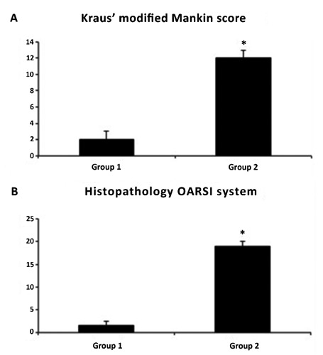
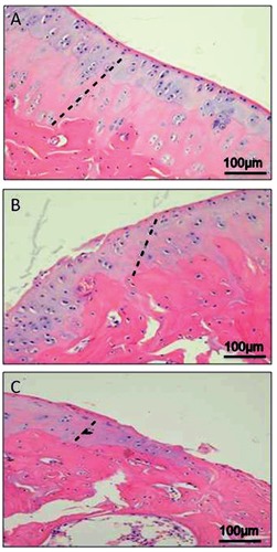
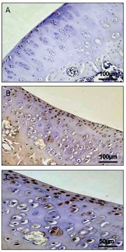
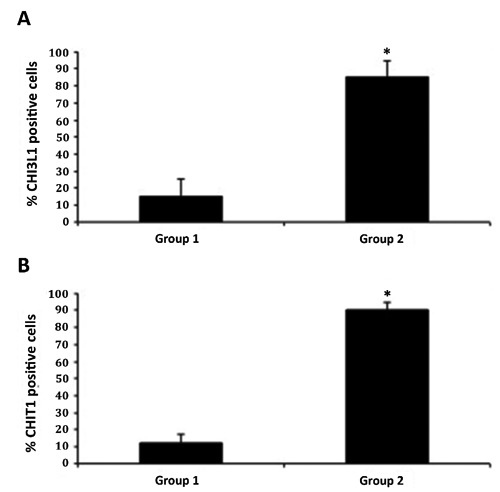
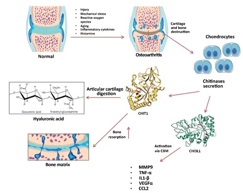
Osteoarthritis is a degenerative joint disease, which affects millions of people around the world. It occurs when the protective cartilage at the end of bones wears over time, leading to loss of flexibility of the joint, pain and stiffness. The cause of osteoarthritis is unknown, but its development is associated with different factors, such as metabolic, genetic, mechanical and inflammatory ones. In recent years the biological role of chitinases has been studied in relation to different inflammatory diseases and more in particular the elevated levels of human cartilage glycoprotein 39 (CHI3L1) and chitotriosidase (CHIT1) have been reported in a variety of diseases including chronic inflammation and degenerative disorders. The aim of this study was to investigate, by immunohistochemistry, the distribution of CHI3L1 and CHIT1 in osteoarthritic and normal rat articular cartilage, to discover their potential role in the development of this disease. The hypothesis was that the expression of chitinases could increase in OA disease. Immunohistochemical analysis showed that CHI3L1 and CHIT1 staining was very strong in osteoarthritic cartilage, especially in the superficial areas of the cartilage most exposed to mechanical load, while it was weak or absent in normal cartilage. These findings suggest that these two chitinases could be functionally associated with the development of osteoarthritis and could be used as markers, so in the future they could have a role in the daily clinical practice to stage the severity of the disease. However, the longer-term in vivo and in vitro studies are needed to understand the exact mechanism of these molecules, their receptors and activities on cartilage tissue.
Conflict of interest statement
Conflict of interests: the authors declare no conflict of interests.
Figures

References
-
- Musumeci G, Loreto C, Carnazza ML, Strehin I, Elisseeff J.OA cartilage derived chondrocytes encapsulated in poly(ethylene glycol) diacrylate (PEGDA) for the evaluation of cartilage restoration and apoptosis in an in vitro model. Histol Histopathol 2011;26:1265-78 - PubMed
-
- Musumeci G, Loreto C, Carnazza ML, Martinez G.Characterization of apoptosis in articular cartilage derived from the knee joints of patients with osteoarthritis. Knee Surg Sports Traumatol Arthrosc 2011;19: 307-13 - PubMed
-
- Musumeci G, Leonardi R, Carnazza ML, Cardile V, Pichler K, Weinberg AM, et al. Aquaporin 1 (AQP1) expression in experimentally induced osteoarthritic knee menisci: an in vivo and in vitro study. Tissue Cell 2013;45:145-52 - PubMed
-
- Musumeci G, Loreto C, Imbesi R, Trovato FM, Di Giunta A, Lombardo C, et al. Advantages of exercise in rehabilitation, treatment and prevention of altered morphological features in knee osteoarthritis. A narrative review. Histol Histopathol 2014; 29:707-19 - PubMed
Publication types
MeSH terms
Substances
LinkOut - more resources
Full Text Sources
Other Literature Sources
Medical
Miscellaneous
