

Non-invasive Foetal ECG - a Comparable Alternative to the Doppler CTG?
- PMID: 25308981
- PMCID: PMC4168331
- DOI: 10.1055/s-0031-1298329
Non-invasive Foetal ECG - a Comparable Alternative to the Doppler CTG?
Abstract
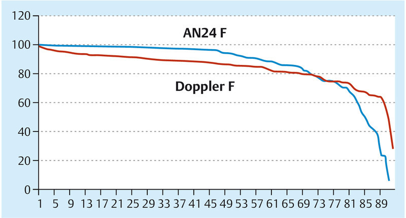
This review discusses the alternative of using the non-invasive foetal ECG compared with the conventionally used Doppler CTG. Non-invasive abdominal electrocardiograms (ECG) have been approved for clinical routine since 2008; subsequently they were also approved for antepartum and subpartum procedures. The first study results have been published. Non-invasive foetal ECG is especially indicated during early pregnancy, while the Doppler CTG is recommended for the vernix period. Beyond the vernix period no difference has been recorded in the success rate of either approach. The foetal ECG signal quality is independent of the BMI, whereas the success rate of the Doppler CTG is diminished with an increased BMI. During the first stage of labour, non-invasive foetal ECG demonstrates better signal quality; however during the second stage of labour no difference has been identified between the methods.
In diesem Review soll die Alternative einer fetalen EKG-Überwachung mit der bisher üblichen CTG-Überwachung verglichen werden. Seit 2008 gibt es ein für die klinische Routine zugelassenes, nicht invasives abdominales Elektrokardiogramm-(EKG-)Gerät, das inzwischen antepartal und unter der Geburt zugelassen ist. Erste Studienergebnisse sind publiziert. Das nicht invasive EKG ist besonders in den frühen Schwangerschaftswochen indiziert, während in der Vernix-Periode das CTG überlegen ist. Nach der Vernix-Periode zeigt sich kein Unterschied in der Erfolgsrate. Die Signalqualität des fetalen EKGs ist vom BMI unabhängig, während das CTG bei einem erhöhten BMI eine schlechtere Signalqualität zeigt. In der Eröffnungsperiode zeigt das EKG eine bessere Signalqualität als das CTG, in der Austreibungsperiode weisen beide Verfahren keine Unterschiede hinsichtlich der Signalqualität auf.
Keywords: Doppler CTG; fetal ECG; obstetrics.
Conflict of interest statement
Figures
Similar articles
-
Prenatal Foetal Non-invasive ECG instead of Doppler CTG - A Better Alternative?Geburtshilfe Frauenheilkd. 2012 Jul;72(7):630-633. doi: 10.1055/s-0032-1315012. Geburtshilfe Frauenheilkd. 2012. PMID: 25278624 Free PMC article.
-
[Foetal electrocardiography (ECG) is an alternative to Doppler ultrasound cardiotocogram (CTG) for antenatal assessment of foetal well-being--preliminary results].Z Geburtshilfe Neonatol. 2008 Dec;212(6):226-9. doi: 10.1055/s-0028-1098718. Epub 2008 Dec 12. Z Geburtshilfe Neonatol. 2008. PMID: 19085740 German.
-
Intrapartum signal quality with external fetal heart rate monitoring: a two way trial of external Doppler CTG ultrasound and the abdominal fetal electrocardiogram.Arch Gynecol Obstet. 2012 Nov;286(5):1103-7. doi: 10.1007/s00404-012-2413-4. Epub 2012 Jun 20. Arch Gynecol Obstet. 2012. PMID: 22714064
-
Foetal scalp blood sampling during labour for pH and lactate measurements.Best Pract Res Clin Obstet Gynaecol. 2016 Jan;30:62-7. doi: 10.1016/j.bpobgyn.2015.05.006. Epub 2015 Jul 8. Best Pract Res Clin Obstet Gynaecol. 2016. PMID: 26253238 Review.
-
[Intrapartum foetal monitoring: from stethoscope to ST analysis of the ECG].Ned Tijdschr Geneeskd. 2009;153:B259. Ned Tijdschr Geneeskd. 2009. PMID: 19785820 Review. Dutch.
References
-
- Shewa A, Hacker T W, Nuovo J. Interpretation of the electronic fetal heart rate during labour. Am Family Phys. 1999;59:2507–2512. - PubMed
-
- Neilson D R, Freeman R K, Mangan S. Signal ambiguity resulting in unexpected outcome with external fetal heart rate monitoring. AJOG. 2008;6:717–724. - PubMed
-
- Amer-Wahlin I, Hellsten C, Norén H. et al.Cardiotocography only versus cardiotocography plus ST analysis of fetal electrocardiogram for intrapartum fetal monitoring: a Swedish randomised controlled trial. Lancet. 2001;358:534–538. - PubMed
-
- Strachan B K, van Wijngaarden W J, Sahota D. et al.Cardiotocography only versus cardiotocography plus PR-interval analysis in intrapartum surveillance: a randomised, multicentre trial. Lancet. 2000;355:456–459. - PubMed
-
- Strachan K B, Sahota D S, van Wijngaarden W J. et al.Computerised analysis of the fetal heart rate and relation to academia at delivery. Br J Obstet Gynaecol. 2001;108:848–852. - PubMed
LinkOut - more resources
Full Text Sources