. 2014 Sep 24:6:258.
doi: 10.3389/fnagi.2014.00258. eCollection 2014.

Astrocytosis in parkinsonism: considering tripartite striatal synapses in physiopathology?

Affiliations

Astrocytosis in parkinsonism: considering tripartite striatal synapses in physiopathology?

Giselle Charron et al. Front Aging Neurosci. .

Abstract

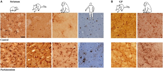
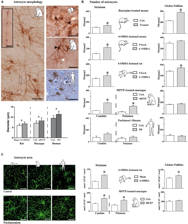
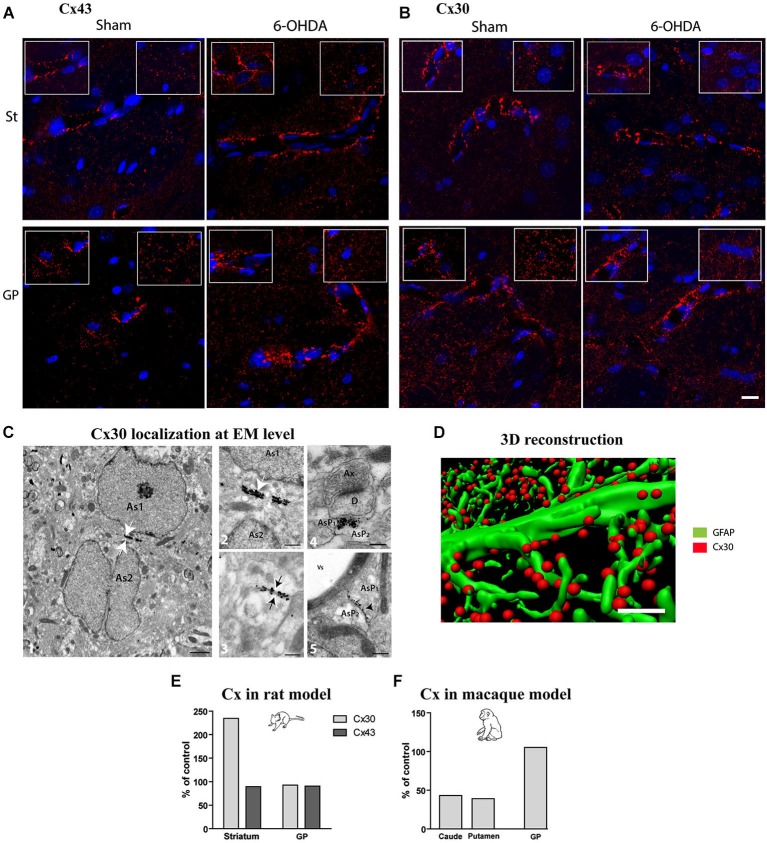
The current concept of basal ganglia organization and function in physiological and pathophysiological conditions excludes the most numerous cells in the brain, i.e., the astrocytes, present with a ratio of 10:1 neuron. Their role in neurodegenerative condition such as Parkinson's disease (PD) remains to be elucidated. Before embarking into physiological investigations of the yet-to-be-identified "tripartite" synapses in the basal ganglia in general and the striatum in particular, we therefore characterized anatomically the PD-related modifications in astrocytic morphology, the changes in astrocytic network connections and the consequences on the spatial relationship between astrocytic processes and asymmetric synapses in normal and PD-like conditions in experimental and human PD. Our results unravel a dramatic regulation of striatal astrocytosis supporting the hypothesis of a key role in (dys) regulating corticostriatal transmission. Astrocytes and their various properties might thus represent a therapeutic target in PD.

Keywords: astrocyte; dopamine; human; immunohistochemistry; medium spiny neuron; monkey; mouse; rat.


Figures

Figure 1
Figure 1
Astrocytic reaction to dopamine depletion in striatum and GP of animal models of PD and parkinsonian patients. Dopamine depletion increase GFAP-S100β immunolabeing in striatum (A) and GP (B) of reserpine mouse, 6-OHDA rat, MPTP non-human primate models of PD and PD patients (scale bar = 30 μm).
Figure 2
Figure 2
Morphological and quantitative analysis of astrocytic reaction to dopamine depletion in striatum and GP of animal models of PD and parkinsonian patients. (A) GFAP-S100β immunostaining enables demonstrating significant increase in astrocyte soma diameter in the rat and monkey models of PD (n = 4 sham and 4 6-OHDA rats; n = 3 control and 3 MPTP monkeys; mean ± SEM) (scale bar = 30 μm but in inset, 10 μm). Primates show two level of immunoreactivity indicated by a black arrowhead for intense labeling and a white arrowhead for moderate staining. (B) Quantitative analysis of astrocyte number in striatum and GP in controls and in animal models of PD (n = 4 reserpine mouse, n = 5 6-OHDA mouse, n = 4 6-OHDA rat, n = 3 MPTP non-human primate) as well as in parkinsonian patients (n = 3) showing increase in astrocyte number. * denotes a significant difference (Student t-test; p < 0.05). (C) Area analysis of astrocytic processes after GFAP immunostaining and 3D reconstruction. Left: representative examples of GFAP immunostaining in the striatum of 6-OHDA rat and MPTP monkey models of PD as well as in PD patients (Scale bar = 30 μm). Right: Astrocytic area significantly increases in the striatum of 6-OHDA rat (n = 4) and MPTP monkey (n = 3) models of PD. * denotes a significant difference (Student t-test ; p < 0.05).
Figure 3
Figure 3
Qualitative and quantitative assessment of connexin 30 and 43 expression in rat and monkey models of PD. Connexin 43 (A) and 30 (B) distribution at cellular level in the striatum (top row) and GP (bottom row) of sham and 6-OHDA rat model of PD. Images correspond to the Z projection of six deconvolved 1 μm optical sections through section, red staining for Cx and blue for nucleus. Insets point out representative Cx immunolabeling around vessels (left) or in neuropil at distance from vessels (right) (scale bar = 10 μm). (C) 1-Representative electron micrographs showing Cx30 localization (scale bar = 1 μm); 2-symmetrically labeled gap junction between two astrocytic soma (white arrow; As = astrocyte) (scale bar = 0.5 μm) ; 3- symmetrically labeled gap junction between two astrocytic processes (black arrow; AsP = astrocytic process) (scale bar = 0.5 μm); 4-symmetrically labeled gap junction between two astrocytic processes close to asymmetrical synapse (scale bar = 0.5 μm); 5-asymmetrical labeled gap junction between two astrocytic processes (scale bar = 0.5 μm). (D) Illustration of quantification methodology of connexin based on double immunolabeling. GFAP immunostaining appears in green while Cx 30 immunostaining appears in red (scale bar = 2 μm). (E) Connexin Cx30 and Cx43 quantification in the 6-OHDA rat and (F) in MPTP monkey models of PD expressed in percentage of levels measured in control animals.
Figure 4
Figure 4
Astrocytes further contacts asymmetric striatal synapses after dopamine depletion. (A) Electron micrograph of GLT1 immunolabeled striatal astrocytes colored in green (pseudocolor image NHI Image J) (scale bar = 2 μm). (B) GLT1-positive astrocytic processes surrounding (asterisk) or not surrounding asymmetric striatal synapses (arrowhead) (scale bar = 0.5 μm). (C) A significant decrease in the number of asymmetric synapses is observed in 6-OHDA rat striatum compared with control (n = 4 animals per group; Student t-test; p < 0.05). (D) Such decrease however decomposes in a significant increase in GLT1+ asymmetric synapses and a dramatic decrease in the number of GLT1− asymmetric synapses in the 6-OHDA rat compared to controls (n = 4 animals per group; one-way ANOVA followed by Bonferroni; p < 0.05).

References

    1. Ball K. K., Gandhi G. K., Thrash J., Cruz N. F., Dienel G. A. (2007). Astrocytic connexin distributions and rapid, extensive dye transfer via gap junctions in the inferior colliculus: implications for [(14)C]glucose metabolite trafficking. J. Neurosci. Res. 85, 3267–3283 10.1002/jnr.21376 - DOI - PMC - PubMed
    1. Berthet A., Bezard E., Porras G., Fasano S., Barroso-Chinea P., Dehay B., et al. (2012). L-DOPA impairs proteasome activity in parkinsonism through D1 dopamine receptor. J. Neurosci. 32, 681–691 10.1523/JNEUROSCI.1541-11.2012 - DOI - PMC - PubMed
    1. Berthet A., Porras G., Doudnikoff E., Stark H., Cador M., Bezard E., et al. (2009). Pharmacological analysis demonstrates dramatic alteration of D1 dopamine receptor neuronal distribution in the rat analog of L-DOPA-induced dyskinesia. J. Neurosci. 29, 4829–4835 10.1523/jneurosci.5884-08.2009 - DOI - PMC - PubMed
    1. Boyes B. E., Kim S. U., Lee V., Sung S. C. (1986). Immunohistochemical co-localization of S-100b and the glial fibrillary acidic protein in rat brain. Neuroscience 17, 857–865 10.1016/0306-4522(86)90050-3 - DOI - PubMed
    1. Braak H., Sastre M., Del Tredici K. (2007). Development of alpha-synuclein immunoreactive astrocytes in the forebrain parallels stages of intraneuronal pathology in sporadic Parkinson’s disease. Acta Neuropathol. 114, 231–241 10.1007/s00401-007-0244-3 - DOI - PubMed

LinkOut - more resources