Review
. 2014 Sep 11:5:452.
doi: 10.3389/fpls.2014.00452. eCollection 2014.

The beginning of a seed: regulatory mechanisms of double fertilization

Affiliations
Review

The beginning of a seed: regulatory mechanisms of double fertilization

Andrea Bleckmann et al. Front Plant Sci. .

Abstract

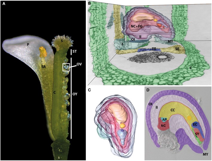
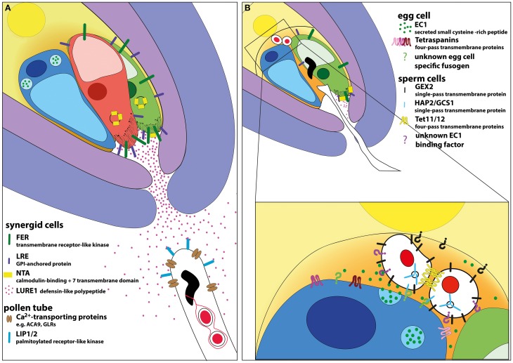
THE LAUNCH OF SEED DEVELOPMENT IN FLOWERING PLANTS (ANGIOSPERMS) IS INITIATED BY THE PROCESS OF DOUBLE FERTILIZATION: two male gametes (sperm cells) fuse with two female gametes (egg and central cell) to form the precursor cells of the two major seed components, the embryo and endosperm, respectively. The immobile sperm cells are delivered by the pollen tube toward the ovule harboring the female gametophyte by species-specific pollen tube guidance and attraction mechanisms. After pollen tube burst inside the female gametophyte, the two sperm cells fuse with the egg and central cell initiating seed development. The fertilized central cell forms the endosperm while the fertilized egg cell, the zygote, will form the actual embryo and suspensor. The latter structure connects the embryo with the sporophytic maternal tissues of the developing seed. The underlying mechanisms of double fertilization are tightly regulated to ensure delivery of functional sperm cells and the formation of both, a functional zygote and endosperm. In this review we will discuss the current state of knowledge about the processes of directed pollen tube growth and its communication with the synergid cells resulting in pollen tube burst, the interaction of the four gametes leading to cell fusion and finally discuss mechanisms how flowering plants prevent multiple sperm cell entry (polyspermy) to maximize their reproductive success.

Keywords: cell fusion; fertilization; gamete interaction; ovule; pollen tube; polyspermy; signaling.


Figures

Figure 1
Figure 1
The female gametophyte is deeply imbedded inside the female flower organs. (A) Dissected and reconstructed Arabidopsis flower. One of four petals (P) and one of six stamina (SA) are shown. They surround the pistil, which represents the female flower organ. It can be dissected into three parts. The upper part contains the papilla cells and forms the stigma (S), which is connected to the ovary (OY) by the style (ST). The ovary is formed by two fused carpels (C), which harbor two rows of ovules (OV). A side view (B) and front view (C) of a 3D-remodeled ovule reconstructed from toluidine blue stained single, successive ultra-thin sections of a dissected pistil. See Supplemental Movie 1 for whole series of sections. The ovule is connected to the septum (SE, yellow) containing the transmitting tract (TT, blue) by the funiculus (F, petrol) and surrounded by the carpel tissue (C) (green). A 3D-model of a dissected ovule shown from various angles is shown in Supplemental Movie 2. The mature female gametophyte cells (FG) and the nucellus tissue (NC) are surrounded by the outer (OI) and inner integuments (II) (OI, blue; II, purple). The vacuole and nucleus of the different female gametophyte cells showed highest contrast and are therefore shown individually. Near to the micropyle (MY), the two nuclei of the two synergid cells (SY) are shown in red and green. The egg cell, indicated by EC in (D), has a comparably large vacuole (light blue) and its nucleus (blue) is located at its chalazal pole. The center of the female gametophyte is filled by the vacuole (light yellow) of the central cell, indicated by CC in (D), and its homo-diploid nucleus (yellow). The three degenerating antipodal cells, indicated by AP in turquoise color in (D) at the chalazal pole are not highlighted. (D) DIC microscopic image of a mature female gametophyte surrounded by the maternal sporophytic tissues of the ovule. The cell types and tissues are artificially colored as shown in (B,C). At full maturity the nucellus cell (NC) layer surrounding the developing embryo sac is flattened between inner integument (II) and female gametophyte cells.
Figure 2
Figure 2
Model of signaling events during micropylar pollen tube attraction and double fertilization in Arabidopsis. (A) The micropylar opening of the ovule is formed by the inner and outer integuments. The female gametophyte is “naked” at its micropylar pole containing one egg (colored in blue) and two synergid cells (colored in green and red) representing the egg apparatus. The central cell surrounds the egg apparatus. The synergid cells are the main sources of pollen tube attractants. Among other components, they secrete LURE peptides, which bind to pollen expressed LIP1/2 receptors thus directing pollen tube growth. Calcium transporters are involved in pollen tube growth control. The plasma membranes of synergid cells harbor a high concentration of receptors like FER and LRE, especially in the region of the filiform apparatus. Upon pollen tube perception NTA is relocated to the plasma membrane by FER activity likely regulated by Ca2+ oscillations inside the synergid cells. (B) The pollen tube bursts when it reaches a certain point beyond the filiform apparatus and releases its cytoplasmic contents including the two sperm cells. Pollen tube burst depends on the presence and activation of FER-, LER-, NTA-, and VDD-dependent signaling cascades culminating in the death of the receptive synergid cell (indicated by diffusing red color). Released sperm cells are located at the gamete fusion side, between the two female gametes. The two sperm cells are connected to each other, likely involving tetraspanins. The male gametes adhere to female gametes by GEX2 located at their surface. After activation, the egg cell secrets EC1 leading to sperm cell activation and HAP2/GCS1 localization to the plasma membrane. HAP2/GCS1 and tetraspanins at the surface of gametes may be involved in mediating membrane fusion. Unknown egg and central cell-specific fusogenic proteins as well as EC1 receptor are indicated by question marks in green, black, and purple, respectively.

References

    1. Alandete-Saez M., Ron M., McCormick S. (2008). GEX3, expressed in the male gametophyte and in the egg cell of Arabidopsis thaliana, is essential for micropylar pollen tube guidance and plays a role during early embryogenesis. Mol. Plant 1, 586–598 10.1093/mp/ssn015 - DOI - PubMed
    1. Amien S., Kliwer I., Márton M. L., Debener T., Geiger D., Becker D., et al. (2010). Defensin-like ZmES4 mediates pollen tube burst in maize via opening of the potassium channel KZM1. PLoS Biol. 8:e1000388 10.1371/journal.pbio.1000388 - DOI - PMC - PubMed
    1. Aw S. J., Hamamura Y., Chen Z., Schnittger A., Berger F. (2010). Sperm entry is sufficient to trigger division of the central cell but the paternal genome is required for endosperm development in Arabidopsis. Development 137, 2683–2690 10.1242/dev.052928 - DOI - PubMed
    1. Barcaccia G., Albertini E. (2013). Apomixis in plant reproduction: a novel perspective on an old dilemma. Plant Reprod. 26, 159–179 10.1007/s00497-013-0222-y - DOI - PMC - PubMed
    1. Bayer M., Nawy T., Giglione C., Galli M., Meinnel T., Lukowitz W. (2009). Paternal control of embryonic patterning in Arabidopsis thaliana. Science 323, 1485–1488 10.1126/science.1167784 - DOI - PubMed