. 2014 Oct 1:6:21.
doi: 10.1186/2045-824X-6-21. eCollection 2014.

Previously differentiated medial vascular smooth muscle cells contribute to neointima formation following vascular injury

Affiliations

Previously differentiated medial vascular smooth muscle cells contribute to neointima formation following vascular injury

Brian Paul Herring et al. Vasc Cell. .

Abstract

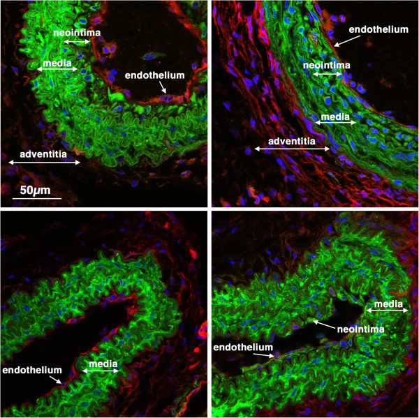
Background: The origins of neointimal smooth muscle cells that arise following vascular injury remains controversial. Studies have suggested that these cells may arise from previously differentiated medial vascular smooth muscle cells, resident stem cells or blood born progenitors. In the current study we examined the contribution of the previously differentiated vascular smooth muscle cells to the neointima that forms following carotid artery ligation.

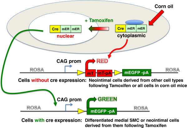
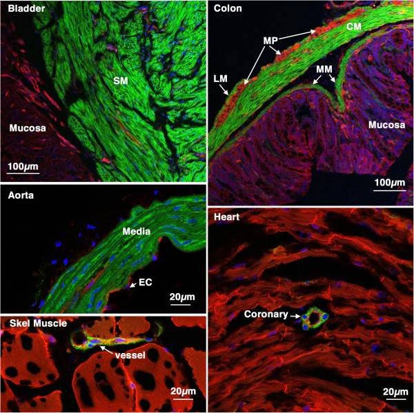
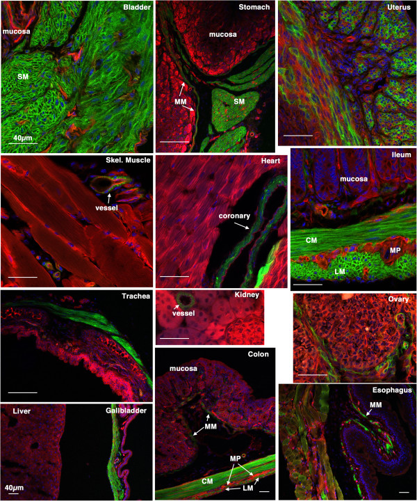
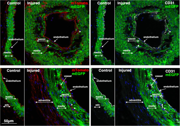
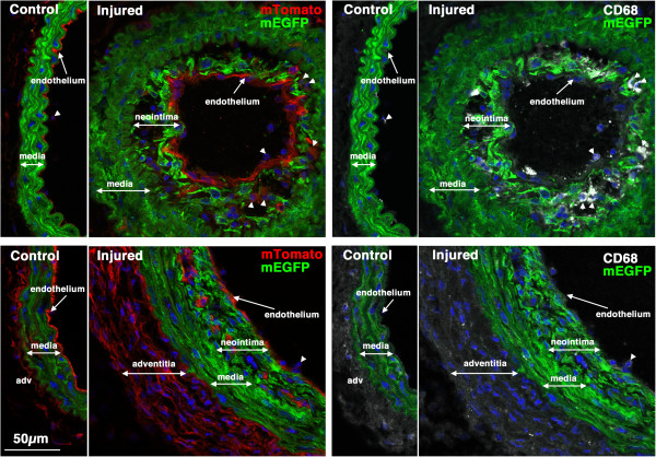
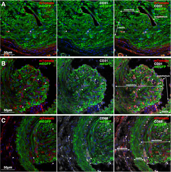
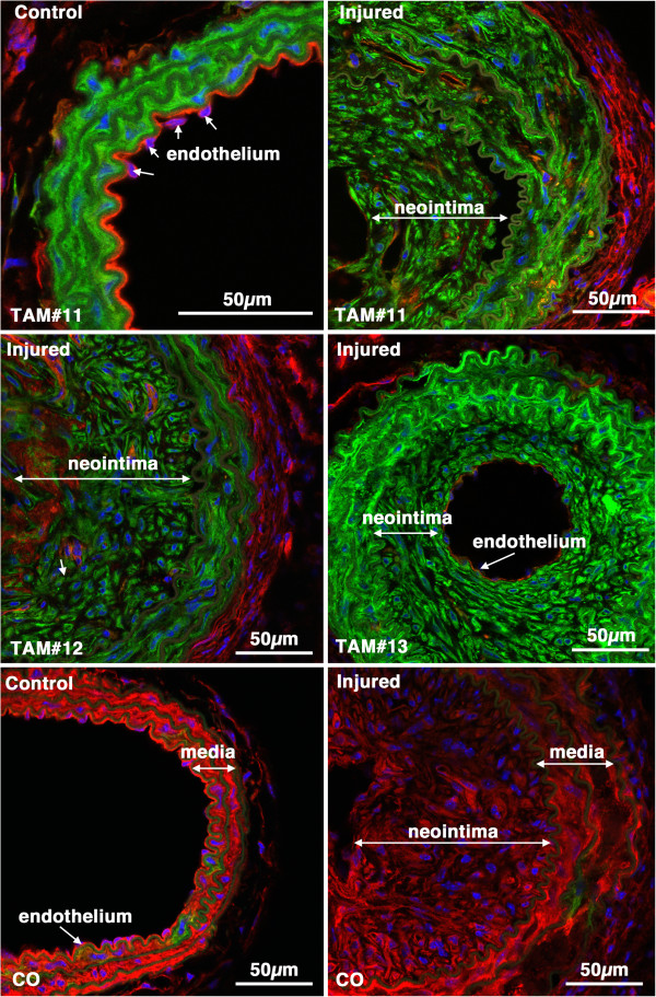
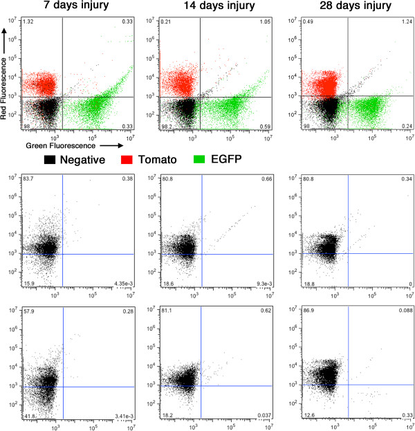
Methods: We utilized transgenic mice harboring a cre recombinase-dependent reporter gene (mTmG). These mice express membrane targeted tandem dimer Tomato (mTomato) prior to cre-mediated excision and membrane targeted EGFP (mEGFP) following excision. The mTmG mice were crossed with transgenic mice expressing either smooth muscle myosin heavy chain (Myh11) or smooth muscle α-actin (Acta2) driven tamoxifen regulated cre recombinase. Following treatment of adult mice with tamoxifen these mice express mEGFP exclusively in differentiated smooth muscle cells. Subsequently vascular injury was induced in the mice by carotid artery ligation and the contribution of mEGFP positive cells to the neointima determined.

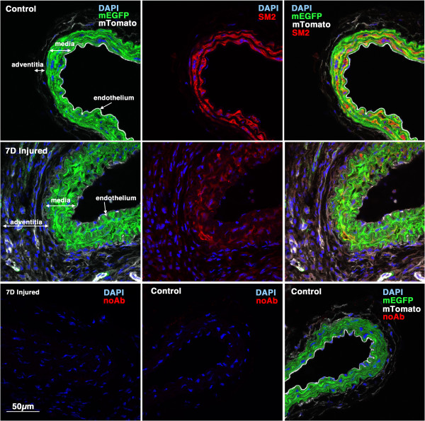
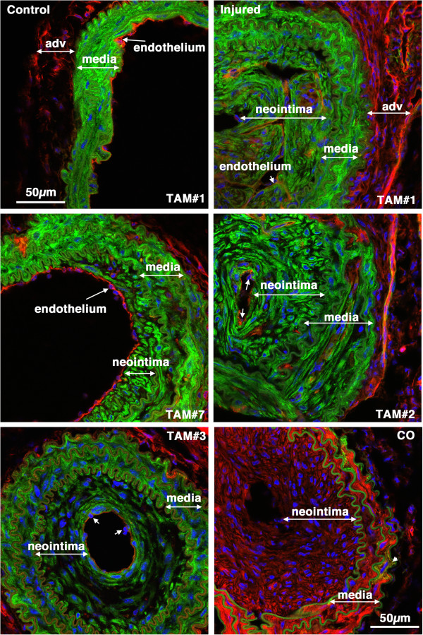
Results: Analysis of the cellular composition of the neointima that forms following injury revealed that mEGFP positive cells derived from either Mhy11 or Acta2 tagged medial vascular smooth muscle cells contribute to the majority of neointima formation (79 ± 17% and 81 ± 12%, respectively).

Conclusion: These data demonstrate that the majority of the neointima that forms following carotid ligation is derived from previously differentiated medial vascular smooth muscle cells.

Keywords: Neointima; Smooth muscle myosin; Smooth muscle α-actin; Vascular smooth muscle.


Figures

Figure 1
Figure 1
Schematic representation of the mTmG reporter strain. The mTmG reporter mice (Jackson strain: B6.129 (Cg)-Gt(ROSA)26Sortm4(ACTB-tdTomato,-EGFP)Luo/J ) contain a single copy of the transgene integrated into the ROSA 26 locus. The transgene cassette is comprised of a chimeric CMV, β-actin promoter driving the expression of a floxed membrane localized Tomato tandem dimer. Following cre-mediated excision the mTomato transgene is removed such that the CAG promoter now drives expression of membrane localized EGFP. The reporter mice were crossed with transgenic mice that express a tamoxifen regulated cre recombinase directed to smooth muscle cells by either the Myh11 promoter or the Acta2 promoter. Double heterozygous transgenic mice were used for all experiments.
Figure 2
Figure 2
Tissue specificity of Myh11 cre ER(T2) activity. 5-week old Myh11 creER(T2)-/+ mTmG-/+ mice were treated with tamoxifen (1 mg,IP) once a day for 5 days. 6 weeks later tissues were harvested and analyzed by confocal microscopy as described in ‘Methods’. A strong mEGFP signal can be seen only in smooth muscle cells of all the tissues examined including, bladder, colon, aorta, skeletal muscle and heart. No cre activity was detected in other cell types including skeletal muscle cells cardiac myocytes, endothelial cells or mucosal epithelial cells. SM-smooth muscle, LM-longitudinal muscle, CM-circular muscle, MP-myenteric plexus, MM-muscularis mucosa, EC-endothelial cells.
Figure 3
Figure 3
Tissue specificity of Acta2 cre ER(T2) activity. 5-week old Acta2 creER(T2)-/+ mTmG-/+ mice were treated with tamoxifen (1 mg,IP) once a day for 5 days. 2 weeks following the last tamoxifen injection the left carotid artery was ligated. 4 weeks later tissues were harvested and analyzed by confocal microscopy as described in ‘Methods’. A strong mEGFP signal can be seen only in smooth muscle cells of all the tissues examined. No cre activity was detected in other cell types including skeletal muscle cells, cardiac myocytes, endothelial cells, mucosal epithelial cells and hepatocytes. A heterogeneous staining was observed in the uterus suggesting that cre was not active in all the uterine smooth muscle cells during the period in which the mice were treated with tamoxifen. The patchwork mEGFP expression observed in the esophagus reflects the mixed skeletal/smooth muscle lineage of cells within the wall of this portion of the esophagus. SM-smooth muscle, LM-longitudinal muscle, CM-circular muscle, MP-myenteric plexus, MM-muscularis mucosa. Scale bars represent 40 μm in all panels.
Figure 4
Figure 4
Myh11 expression is down-regulated 7 days following injury. 5-week old male Myh11 creER(T2)-/+ mTmG-/+ mice were treated with tamoxifen (1 mg,IP) once a day for 5 days. Two weeks following the last tamoxifen injection the left carotid artery was ligated and tissues were harvested 7 days later. 8 μm cryosections obtained from injured and contralateral control arteries were analyzed for expression of myh11 (using an anti-SM2 antibody) (red) and mEGFP (green) and mTomato (white). Control and injured sections are shown at identical exposures. Images shown are representative of those obtained from 3 different mice. In the lower panels the SM2 antibody was omitted and the samples otherwise processed identically to those shown in the upper panels. All images are shown at the same magnification with the scale bar shown in the bottom left panel representing 50 μm.
Figure 5
Figure 5
Previously differentiated VSMC contribute to early neointima formation. 5-week old male Myh11 creER(T2)-/+ mTmG-/+ mice were treated with tamoxifen (1 mg,IP) once a day for 5 days. Two weeks following the last tamoxifen injection the left carotid artery was ligated and tissues were harvested 14 days later. Expression of mTomato (red) and mEGFP (green) were visualized in sections obtained from 4 different mice. Nuclei were visualized by staining with DAPI (blue). All images are shown at the same magnification with the scale bar shown in the top left panel representing 50 μm.
Figure 6
Figure 6
CD31 positive endothelial cells can be seen within early developing neointima. Sections from two of the mice shown in Figure  5 were stained with antibodies to CD31 (white). Left panels show mTomato (red)/mEGFP (green) co-stained sections and in the right panels are the same sections visualized for mEGFP (green) and CD31(white). Nuclei were visualized by staining with DAPI (blue). All images are shown at the same magnification with the scale bar shown in the bottom left panel representing 50 μm.
Figure 7
Figure 7
CD68 positive macrophages/monocytes cells can be seen within early developing neointima in some mice. Sections from the same two mice shown in Figure  6 were stained with antibodies to CD68 (white). Left panels show mTomato (red)/mEGFP (green) co-stained sections and in the right panels are the same sections visualized for mEGFP (green) and CD68(white). CD68-positive cells can be seen within the neointima of vessels from the mouse shown in the upper panels but not in the vessels of the mouse shown in the lower panels. Nuclei were visualized by staining with DAPI (blue). All images are shown at the same magnification with the scale bar shown in the bottom left panel representing 50 μm.
Figure 8
Figure 8
Myh11-tagged medial SMCs give rise to the mature neointima following carotid ligation. Tissues were harvested 28 days following carotid ligation of 5-week old male Myh11 creER(T2)-/+ mTmG-/+ mice that were previously Treated with Tamoxifen (TAM) or corn oil(CO) as indicated. In the upper two panels the control and injured carotid arteries from the same mouse are shown. The neointima, media and adventitia (adv) are indicated. Images are representative of those obtained from 7 tamoxifen treated mice (Mice numbers 1,7,2,3 in Table  1, as indicated). Arrows point to examples of endothelial cell nuclei. The image in the lower right hand panel is of an injured vessel obtained from a corn oil control treated mouse. The arrow-head in this image points to an mEGFP positive cell within the medial layer. All images are shown at the same magnification with the scale bars representing 50 μm.
Figure 9
Figure 9
CD31 and CD68 positive cells in mature neointimal lesions. Images shown are examples of mature lesions seen in 2 different Myh11 creER(T2)-/+ mTmG-/+ mice 28 days following ligation (Mouse 4(A) and 6(B, C); Table  1). A, B. Sections were immuno-stained for the endothelial/platelet marker CD31. Images shown are merged images showing the mTomato (red), mEGFP (green) and DAPI (blue); CD31 (white), mEGFP (green) and DAPI (blue) or mTomato, mEGFP, CD31 and DAPI channels, as indicated. Sections obtained from the mouse shown in panel A exhibited very few CD31 positive cells in the mature neointima; in contrast the lesion shown in panel B exhibited numerous CD31 positive, mTomato positive, mEGFP negative cells in the neointima (examples indicated by arrow heads). C. Sections (serial to those shown in panel B) were immuno-stained for the macrophage/monocyte marker CD68. Images shown are merged images showing the mTomato (red), mEGFP (green) and DAPI (blue); CD68 (white), mEGFP (green) and DAPI (blue) or mTomato, mEGFP, CD68 and DAPI channels, as indicated. Several CD68 positive, mTomato positive, mEGFP negative cells can be seen in the neointima and adventitia (examples indicated by arrow heads). No CD68 positive cells were seen in the neointima of the mouse shown in panel A (data not shown).
Figure 10
Figure 10
Acta2-tagged medial SMCs give rise to the neointima following carotid ligation. Control and injured carotid arteries from Tamoxifen (TAM) or corn oil (CO) treated Acta2 creER(T2)-/+ mTmG-/+ transgenic mice harvested 28 days post ligation (Mice numbers 11, 12 and 13, respectively; Table  1). Images from tamoxifen treated injured vessels are representative of those obtained from 7 mice.
Figure 11
Figure 11
No mEGFP positive circulating cells can be detected following carotid ligation. Blood was harvested from Myh11 creER(T2)-/+ mTmG-/+ mice 7, 14 and 28 days following carotid ligation and subjected to fluorescence activated cell sorting. Cells harvested from a CAG-EGFP mouse were used as positive controls for EGFP expressing cells, those harvested from mTmG reporter mice with no cre transgene as positive controls for mTomato positive cells and those harvested from nontransgenic mice as negative controls. From each mouse a minimum of 10,000 cells were sorted. Upper panels show the distribution of mTomato (red) and mEGFP (green) positive as well as negative (black) cells. Numbers on each graph show the percentage of negative cells in each quadrant. Lower panels show the distribution of cells obtained from duplicate Myh11 creER(T2)-/+ mTmG-/+ mice. In each case the percentage of EGFP-positive cells are less than background negative control levels.

References

    1. Dandre F, Owens GK. Platelet-derived growth factor-BB and Ets-1 transcription factor negatively regulate transcription of multiple smooth muscle cell differentiation marker genes. Am J Physiol Heart Circ Physiol. 2004;286:H2042–H2051. doi: 10.1152/ajpheart.00625.2003. - DOI - PubMed
    1. Hu Y, Davison F, Ludewig B, Erdel M, Mayr M, Url M, Dietrich H, Xu Q. Smooth muscle cells in transplant atherosclerotic lesions are originated from recipients, but not bone marrow progenitor cells. Circulation. 2002;106:1834–1839. doi: 10.1161/01.CIR.0000031333.86845.DD. - DOI - PubMed
    1. Bentzon JF, Sondergaard CS, Kassem M, Falk E. Smooth muscle cells healing atherosclerotic plaque disruptions are of local, not blood, origin in apolipoprotein E knockout mice. Circulation. 2007;116:2053–2061. doi: 10.1161/CIRCULATIONAHA.107.722355. - DOI - PubMed
    1. Iwata H, Manabe I, Fujiu K, Yamamoto T, Takeda N, Eguchi K, Furuya A, Kuro-o M, Sata M, Nagai R. Bone marrow-derived cells contribute to vascular inflammation but do not differentiate into smooth muscle cell lineages. Circulation. 2010;122:2048–2057. doi: 10.1161/CIRCULATIONAHA.110.965202. - DOI - PubMed
    1. Nemenoff RA, Horita H, Ostriker AC, Furgeson SB, Simpson PA, VanPutten V, Crossno J, Offermanns S, Weiser-Evans MC. SDF-1alpha induction in mature smooth muscle cells by inactivation of PTEN is a critical mediator of exacerbated injury-induced neointima formation. Arterioscler Thromb Vasc Biol. 2011;31:1300–1308. doi: 10.1161/ATVBAHA.111.223701. - DOI - PMC - PubMed

LinkOut - more resources