. 2014:2014:679672.
doi: 10.1155/2014/679672. Epub 2014 Sep 17.

Planarians sense simulated microgravity and hypergravity

Affiliations

Planarians sense simulated microgravity and hypergravity

Teresa Adell et al. Biomed Res Int. 2014.

Abstract

Planarians are flatworms, which belong to the phylum Platyhelminthes. They have been a classical subject of study due to their amazing regenerative ability, which relies on the existence of adult totipotent stem cells. Nowadays they are an emerging model system in the field of developmental, regenerative, and stem cell biology. In this study we analyze the effect of a simulated microgravity and a hypergravity environment during the process of planarian regeneration and embryogenesis. We demonstrate that simulated microgravity by means of the random positioning machine (RPM) set at a speed of 60 °/s but not at 10 °/s produces the dead of planarians. Under hypergravity of 3 g and 4 g in a large diameter centrifuge (LDC) planarians can regenerate missing tissues, although a decrease in the proliferation rate is observed. Under 8 g hypergravity small planarian fragments are not able to regenerate. Moreover, we found an effect of gravity alterations in the rate of planarian scission, which is its asexual mode of reproduction. No apparent effects of altered gravity were found during the embryonic development.


Figures

Figure 1
Figure 1
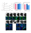
(a) Planarians' regeneration timing. When planarians are cut into head, trunk, and tail pieces, they produce a blastema (unpigmented region at day 2), and the subsequent regeneration of all missing tissues in 10–15 days. The yellow asterisk labels the pharynx. (b) The two environments in which planarians were cultured both with sample and control groups. Animals were included in falcon tubes, filled with planarian water, at a density of 10 animals per falcon.
Figure 2
Figure 2
Results of Experiment 1. Planarians regenerate in 3 g hypergravity but not in the microgravity simulated by the 60°/s RPM. (a) Schedule of Experiment 1. (b) Comparison between sample group and control group in the RPM. At day 13 all RPM animals appeared dead, while controls were regenerated and healthy. (c) Comparison between sample group and control group in the LDC. DAPI staining (green) shows that both groups regenerated properly the missing structures. In both groups animals fissioned during the experiment and thus at day 13 animals at different stages of regeneration are found. In the image, a completely regenerated brain in the anterior region is labelled with white arrows and a regenerating one of a planarian fragment is indicated by a yellow arrowhead. The counting of pH3 positive cells in LDC planarians, grouped according to their morphology after their differential fissioning, indicates that under hypergravity conditions there is a slight decrease in the number of proliferative cells, although it is not statistically significant. Graphs error bars represent standard error of the mean. Data were analyzed by Student's t-test. Differences are considered significant at P < 0.05.
Figure 3
Figure 3
Results of Experiment 2. Planarian trunks can regenerate properly in the RPM at 10°/s and under 4 g and 8 g LDC hypergravity. (a) Schedule of Experiment 2. (b) At day 5 all animals are regenerating properly (see the two black spots in the in vivo image and the brain stained with DAPI, in blue, and anti-synapsin, in green). At day 13 all animals are properly regenerated but RPM 60°/s, which were dead in 87,5% (see the eyes, the brain stained with anti-synapsin and the visual axons stained with anti-VC1). (c) The counting of pH3 positive cells in RPM and LDC planarians at days 5 and 12 of regeneration indicates that under hypergravity there is a significant decrease in the number of proliferative cells at 12 days of regeneration. Graphs error bars represent standard error of the mean. Data were analyzed by Student's t-test. Differences are considered significant at P < 0.05. Orange arrows indicate the regenerating eyes. Yellow arrows indicate the regenerating brain. White arrows indicate the regenerating eyes stained with anti-VC1. Yellow arrowhead points to the optic chiasm.
Figure 4
Figure 4
Hypergravity impairs regeneration of small planarian fragments. General images of the different planarian groups in the LDC conditions at the end of Experiment 2 are shown in the upper panel. Note that at 8 g all planarians are dead. In the lower panel stereoscopic images of planarians show that, while control fragments regenerated properly, head fragments under 4 g conditions present an aberrant morphology. Moreover, no tail fragments were found, which means they died during the experiment.
Figure 5
Figure 5
Simulated alteration of gravity affects planarian scission rate. (a) Scheme of the groups corresponding to Experiment 2 in which scission rates were analyzed. (b) At day 1 several fissioning animals were found (red arrow, in upper images). At day 13 a higher number of small fragments were found in LDC 8 g conditions compared to controls (lower images). (c) Quantification of the fissioned animals at day 1 of the experiment shows that RPM (60°/s) decreases while LDC (8 g) increases it. (d) Quantification of the number of small fissioned fragments in relation to the number of original planarians shows a higher rate of fissioned animals under 4 g hypergravity. In the graphs error bars represent standard error of the mean. Data were analyzed by Student's t-test. **P < 0.001; ***P < 0.001. Differences are considered significant at P < 0.05.
Figure 6
Figure 6
Planarian embryos develop in simulated microgravity (RPM 60°/s) and in hypergravity (3 g) environments. (a) Image of a planarian cocoon and a hatchling planarian. (b) Immunohistochemical analysis of planarian hatchlings corresponding to all conditions tested shows their normal morphology (see the nervous system stained with anti-α-tubulin, in green, and the pharynx and brain stained with DAPI, in blue). White arrows indicate the brain and yellow asterisk indicates the pharynx. (c) Quantification of the pH3 shows no significant differences in the mitotic rates among the different groups. Data were analyzed by Student's t-test. Differences are considered significant at P < 0.05.

References

    1. Saló E, Baguñà J. Regeneration in planarians and other worms: new findings, new tools and new perspectives. Journal of Experimental Zoology. 2002;292(6):528–539. - PubMed
    1. Reddien PW, Alvarado S. Fundamentals of planarian regeneration. Annual Review of Cell and Developmental Biology. 2004;20:725–757. - PubMed
    1. Saló E. The power of regeneration and the stem-cell kingdom: freshwater planarians (Platyhelminthes) BioEssays. 2006;28(5):546–559. - PubMed
    1. Alvarado AS. Planarian regeneration: its end is its beginning. Cell. 2006;124(2):241–245. - PubMed
    1. Handberg-Thorsager M, Fernandez E, Saló E. Stem cells and regeneration in planarians. Frontiers in Bioscience. 2008;13(16):6374–6394. - PubMed

Publication types

LinkOut - more resources