. 2014 Oct 13;9(10):e105311.
doi: 10.1371/journal.pone.0105311. eCollection 2014.

Withanolide A prevents neurodegeneration by modulating hippocampal glutathione biosynthesis during hypoxia

Affiliations

Withanolide A prevents neurodegeneration by modulating hippocampal glutathione biosynthesis during hypoxia

Iswar Baitharu et al. PLoS One. .

Abstract

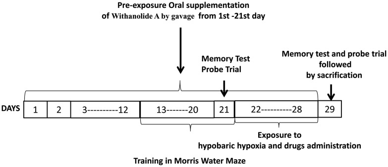
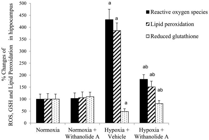
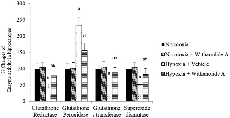
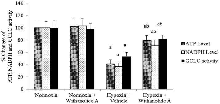
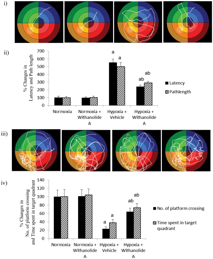
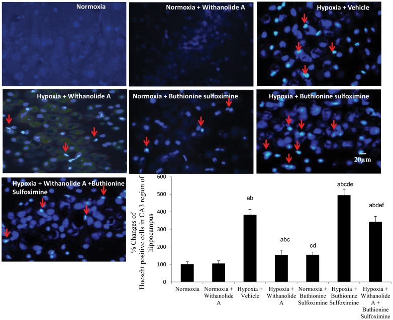
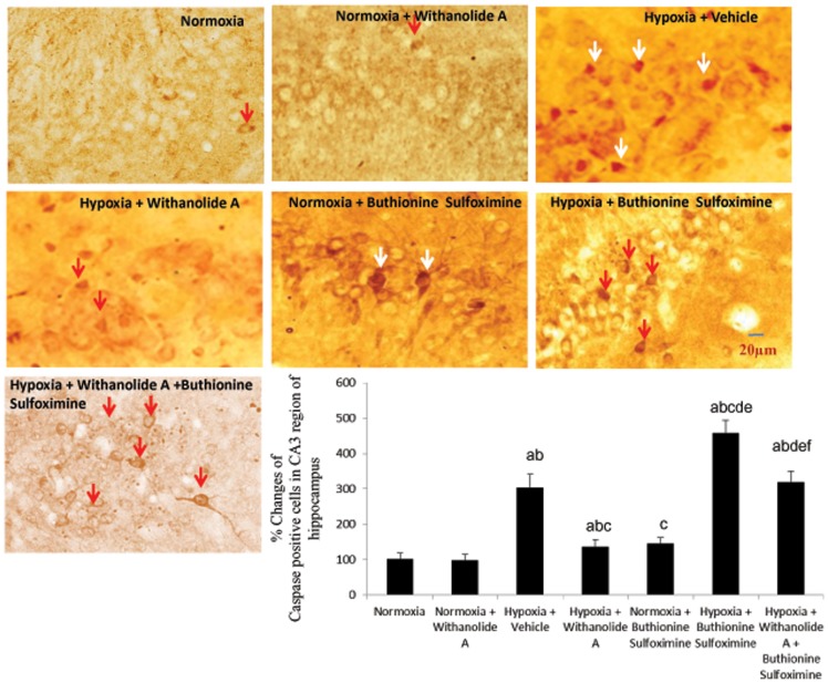
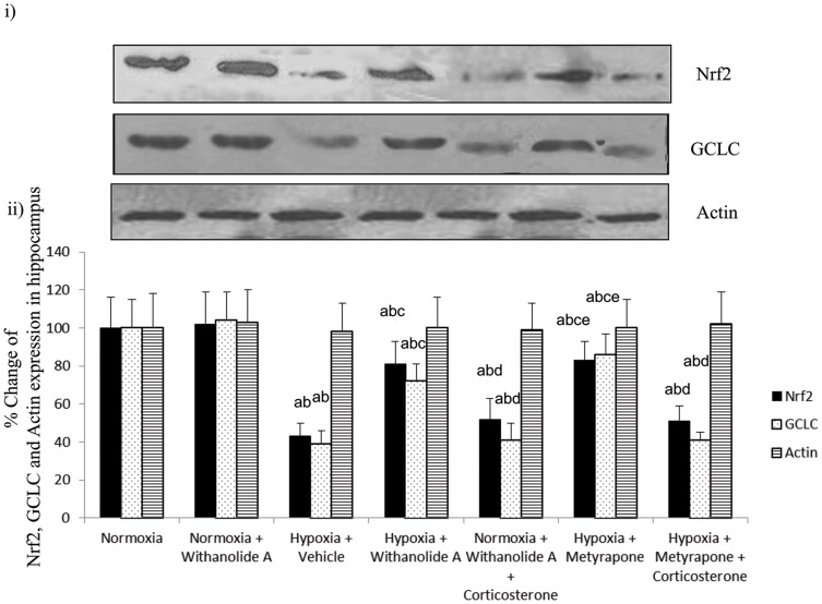
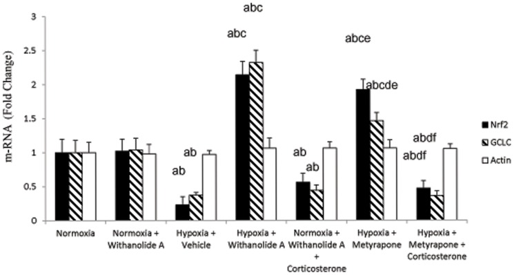
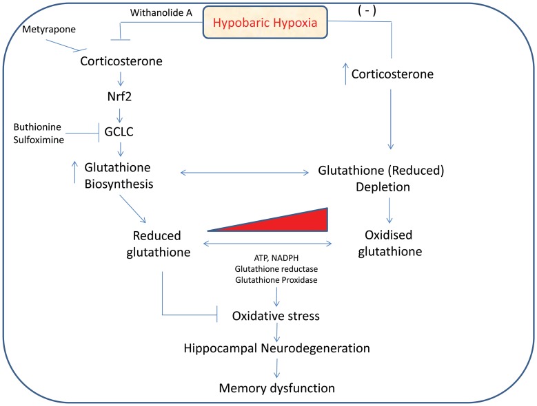
Withania somnifera root extract has been used traditionally in ayurvedic system of medicine as a memory enhancer. Present study explores the ameliorative effect of withanolide A, a major component of withania root extract and its molecular mechanism against hypoxia induced memory impairment. Withanolide A was administered to male Sprague Dawley rats before a period of 21 days pre-exposure and during 07 days of exposure to a simulated altitude of 25,000 ft. Glutathione level and glutathione dependent free radicals scavenging enzyme system, ATP, NADPH level, γ-glutamylcysteinyl ligase (GCLC) activity and oxidative stress markers were assessed in the hippocampus. Expression of apoptotic marker caspase 3 in hippocampus was investigated by immunohistochemistry. Transcriptional alteration and expression of GCLC and Nuclear factor (erythroid-derived 2)-related factor 2 (Nrf2) were investigated by real time PCR and immunoblotting respectively. Exposure to hypobaric hypoxia decreased reduced glutathione (GSH) level and impaired reduced gluatathione dependent free radical scavenging system in hippocampus resulting in elevated oxidative stress. Supplementation of withanolide A during hypoxic exposure increased GSH level, augmented GSH dependent free radicals scavenging system and decreased the number of caspase and hoescht positive cells in hippocampus. While withanolide A reversed hypoxia mediated neurodegeneration, administration of buthionine sulfoximine along with withanolide A blunted its neuroprotective effects. Exogenous administration of corticosterone suppressed Nrf2 and GCLC expression whereas inhibition of corticosterone synthesis upregulated Nrf2 as well as GCLC. Thus present study infers that withanolide A reduces neurodegeneration by restoring hypoxia induced glutathione depletion in hippocampus. Further, Withanolide A increases glutathione biosynthesis in neuronal cells by upregulating GCLC level through Nrf2 pathway in a corticosterone dependenet manner.


Conflict of interest statement

Competing Interests: The authors have declared that no competing interests exist.

Figures

Figure 1
Figure 1. Showing the schedule of the training, probe trial and memory test in Morris Water Maze, supplementation of Withanolide A, administration of drugs and exposure to hypobaric hypoxia.
Figure 2
Figure 2. Effect of Withanolide A on oxidative stress markers.
Administration of Withanolide A decreases the hypoxia induced elevated level of reactive oxygen species, lipid peroxidation and GSH in hippocampus. Data expressed as percentage change taking normoxic value as 100% and represents Mean ± SEM. ‘a’ denotes p≤0.05 vs. when compared to normoxic group and ‘b’ denotes p≤0.05 vs. when compared to 7 days hypoxic group treated with vehicle only.
Figure 3
Figure 3. Effect of Withanolide A on free radical scavenging enzyme system.
Withanolide A administration during hypoxic exposure increases hypoxia induced decreased activity of glutathione reductase, glutathione peroxidase, glutathione s transferase and superoxide dismutase in hippocampus. Data expressed as percentage change taking normoxic value as 100% and represents Mean ± SEM. ‘a’ denotes p≤0.05 vs. when compared to normoxic group and ‘b’ denotes p≤0.05 vs. when compared to 7 days hypoxic group treated with vehicle only.
Figure 4
Figure 4. Restoration of ATP, NADPH and γ-glutamylcysteinyl ligase catalytic subunit activity in hippocampus following Withanolide A during hypoxia.
Hypoxic exposure for 7 days decreases the hippocampal ATP, NADPH and γ-glutamylcysteinyl ligase catalytic subunit activity while administration of Withanolide A increases the ATP, NADPH and γ-glutamylcysteinyl ligase catalytic subunit activity in hippocampus. Data expressed as percentage change taking normoxic value as 100% and represents Mean ± SEM. ‘a’ denotes p≤0.05 vs. when compared to normoxic group and ‘b’ denotes p≤0.05 vs. when compared to 7 days hypoxic group treated with vehicle only.
Figure 5
Figure 5. Amelioration of spatial memory function following Withanolide A administration during hypoxic exposure.
Exposure to hypobaric hypoxia increased (i) path length and (ii) latency during spatial memory test but decreased (iii) number of platform crossing and (iv) time spent in target quadrant during probe trial when was reversed following withanolide A supplementation before and during exposure to hypobaric hypoxia. Data expressed as percentage change taking normoxic value as 100% and represents Mean ± SEM. ‘a’ denotes p≤0.05 vs. when compared to normoxic group and ‘b’ denotes p≤0.05 vs. when compared to 7 days hypoxic group treated with vehicle only.
Figure 6
Figure 6. Modulation of hippocampal endogenous glutathione level by Withanolide A reduces hypoxia induced neurodegeneration.
Withanolide A effectively decreases the number of degenerating neurons caused by hypoxic exposure. However, depletion of glutathione using buthionine sulfoximine during hypoxic exposure increases the number degenerating cells in hippocampus. Co-administration of withanolide A alongwith buthionine sulfoximine attenuates the neuroprotective effect of withanolide A during hypoxia. Data expressed as percentage change taking normoxic value as 100% and represents Mean ± SEM. ‘a’ denotes p≤0.05 vs. when compared to normoxic group. ‘b’ denotes p≤0.05 vs. when compared to normoxia + withanolide A group and ‘c’ denotes p≤0.05 vs. when compared to hypoxia + vehicle group, ‘d’ denotes p≤0.05 vs. when compared to hypobaric hypoxia + withanolide A, ‘e’ denotes p≤0.05 vs. when compared to normoxia + buthionine sulfoximine, and ‘f’ denotes p≤0.05 vs. when compared to hypobaric hypoxia + buthionine sulfoximine.
Figure 7
Figure 7. Withanolide A mediated restoration of endogenous glutathione level reverses hypoxia induced neuronal apoptosis in hippocampus.
Administration of withanolide A prior to and during hypoxic exposure decreases number of apoptotic cells in CA3 region of hippocampus while glutathione depletion using buthionine sulfoximine elevates hypoxic neuronal apoptosis. Co-administration of withanolide A alongwith buthionine sulfoximine during hypoxic exposure enhances hypoxia induced apoptotic cells attenuating the nuroprotective effect of withanolide A. Data expressed as percentage change taking normoxic value as 100% and represents Mean ± SEM ‘a’ denotes p≤0.05 vs. when compared to normoxic group. ‘b’ denotes p≤0.05 vs. when compared to normoxia + withanolide A group and ‘c’ denotes p≤0.05 vs. when compared to hypoxia + vehicle group, ‘d’ denotes p≤0.05 vs. when compared to hypobaric hypoxia + withanolide A, ‘e’ denotes p≤0.05 vs. when compared to normoxia + buthionine sulfoximine, and ‘f” denotes p≤0.05 vs. when compared to hypobaric hypoxia + buthionine sulfoximine.
Figure 8
Figure 8. Withanolide A mediated elevation of hippocampal glutathione during hypoxia is corticosterone dependent.
Withanolide A administration during hypoxic exposure upregulates Nrf2 and GCLC expression in hippocampus. Administration of Withanolide A alongwith exogenous corticosterone supplementation to the normoxic group decrease the Nrf2 as well as GCLC expression while inhibition of corticosterone synthesis using metyrapone reverses hypoxia induced downregulation of both Nrf2 and GCLC level. β-actin was used as a loading control. Data expressed as percentage change taking normoxic value as 100% and represents Mean ± SEM. ‘a’ denotes p≤0.05 vs. when compared to normoxic group. ‘b’ denotes p≤0.05 vs. when compared to normoxia + withanolide A group and ‘c’ denotes p≤0.05 vs. when compared to hypoxia + vehicle group, ‘d’ denotes p≤0.05 vs. when compared to hypobaric hypoxia + withanolide A, ‘e’ denotes p≤0.05 vs. when compared to hypobaric hypoxia + withanolide A + corticosterone and ‘f” denotes p≤0.05 vs. when compared to hypobaric hypoxia + Metyrapone.
Figure 9
Figure 9. Withanolide A mediated transcriptional regulation of Nrf2 and GCLC expression depend on corticosterone signaling in hippocampus.
Withanolide A administration during hypoxic exposure upregulates Nrf2 and GCLC expression in hippocampus. Administration of Withanolide A alongwith exogenous corticosterone supplementation to the normoxic group decrease the Nrf2 as well as GCLC expression while inhibition of corticosterone synthesis using metyrapone during hypoxia reverses hypoxia induced downregulation of both Nrf2 and GCLC level. β-actin was used as a loading control. Data expressed as percentage change taking normoxic value as 100% and represents Mean ± SEM. ‘a’ denotes p≤0.05 vs. when compared to normoxic group. ‘b’ denotes p≤0.05 vs. when compared to normoxia + withanolide A group and ‘c’ denotes p≤0.05 vs. when compared to hypoxia + vehicle group, ‘d’ denotes p≤0.05 vs. when compared to hypobaric hypoxia + withanolide A, ‘e’ denotes p≤0.05 vs. when compared to hypobaric hypoxia + withanolide A + corticosterone and ‘f” denotes p≤0.05 vs. when compared to hypobaric hypoxia + metyrapone.
Figure 10
Figure 10. Schematic diagram showing Withanolide A mediated modulation of glutathione biosynthesis and neuroprotection during hypobaric hypoxia.

References

    1. Bahrke M, Hale BS (1993) Effect of altitude on mood, behavior and cognitive functioning. Sports Med 16: 97–125. - PubMed
    1. Baitharu I, Jain V, Deep SN, Sahu JK, Naik PK, et al. (2013) Exposure to hypobaric hypoxia and reoxygenation induces transient anxiety like behaviour in rat. J Behav Brain Sci 3: 591–602.
    1. Won SJ, Kim DY, Gwag BJ (2002) Cellular and molecular pathways of ischemic neuronal death. J Biochem Mol Biol 35: 67–86. - PubMed
    1. Barker JE, Heales SJR, Cassidy A, Bolanos JP, Land JM, et al. (1996) Depletion of brain glutathione results in a decrease in glutathione reductase activity, an enzyme susceptible to oxidative damage. Brain Res 716: 118–122. - PubMed
    1. Maiti P, Singh SB, Sharma AK, Muthuraju S, Banerjee PK, et al. (2006) Hypobaric hypoxia induces oxidative stress in rat brain. Neurochem International 49: 709–716. - PubMed

Publication types

MeSH terms