. 2014 Oct 13;9(10):e109152.
doi: 10.1371/journal.pone.0109152. eCollection 2014.

Characterization of PHB1 and its role in mitochondrial maturation and yolk platelet degradation during development of Artemia embryos

Affiliations

Characterization of PHB1 and its role in mitochondrial maturation and yolk platelet degradation during development of Artemia embryos

Xiang Ye et al. PLoS One. .

Abstract

Background: To cope with harsh environments, crustaceans such as Artemia produce diapause gastrula embryos (cysts) with suppressed metabolism. Metabolism and development resume during post-diapause development, but the mechanism behind these cellular events remains largely unknown.

Principal finding: Our study investigated the role of prohibitin 1 (PHB1) in metabolic reinitiation during post-diapause development. We found that PHB1 was developmentally regulated via changes in phosphorylation status and localization. Results from RNA interference experiments demonstrated PHB1 to be critical for mitochondrial maturation and yolk degradation during development. In addition, PHB1 was present in yolk platelets, and it underwent ubiquitin-mediated degradation during the proteolysis of yolk protein.

Conclusions/significance: PHB1 has an indispensable role in coordinating mitochondrial maturation and yolk platelet degradation during development in Artemia. This novel function of PHB1 provides new clues to comprehend the roles of PHB1 in metabolism and development.


Conflict of interest statement

Competing Interests: The authors have declared that no competing interests exist.

Figures

Figure 1
Figure 1. Sequence and phylogenetic analysis of ArPHB1.
A, Schematic view of PHB1 architecture. B, Amino acid sequence alignment of ArPHB1 with PHB1 from other species. The sequences used in this alignment and their Genbank/EMBL/DDBJ or SWISS-PROT database accession numbers are listed in Table S2. Gaps inserted to maximize alignment are denoted by hyphens. The amino acid position numbers are shown to the right of the sequences. The phosphorylation sites against which the antibody was generated is marked by P. Conserved domains are marked by lines below sequences (the N-terminal hydrophobic domain by yellow, the PHB domain by black, the coiled-coil domain by blue, and the nuclear export sequence by red). The alignment was performed by MEGA5 using the ClustalW method. C, A phylogenetic tree of the amino acid sequences of prohibitins. The sequences used in this analysis and their GenBank/EMBL/DDBJ or SWISS-PROT database accession numbers are listed in Table S1. The phylogenetic tree was constructed using the Neighbor-joining method. Bootstrap percentage values for 1000 replicate analysis are shown at branching points. The bar at the bottom shows the branch length, and it corresponds to the mean number of differences (0.05) per residue along each branch.
Figure 2
Figure 2. Characterization of ArPHB1 during post-embryonic development.
A, Western blot analysis of PHB1. 0 h, 0 h-incubated cysts; 6 h, 6 h-incubated cysts; 12 h, 12 h-incubated cysts; 24 h, 24 h-incubated nauplius. B, Western blot analysis of phosphorylated PHB1 and PHB1 modifications. The same samples were loaded in B as in A. C, PHB1-75 existed as a non-membrane form of PHB1 in Artemia. Mem, membrane fraction obtained by 20000×g centrifugation after nuclei isolation (800×g, 10 min); Cyto, cytosol fraction obtained after 20000×g centrifugation. PHB1-75 was marked by an asterisk.
Figure 3
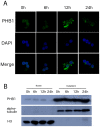
Figure 3. The localization of PHB1 in nuclei during Artemia post-embryonic development.
A, Immunofluorescence analysis of the nucleus. Nuclei from 0, 6, 12, and 24 h-incubated nauplii were isolated and stained with an anti-PHB1 antibody. DNA was stained with DAPI (4,6-diamidino-2-phenylindole dilactate). B, Nuclei from the respective developmental stages were isolated and analyzed by Western blotting (anti-PHB1).
Figure 4
Figure 4. ArPHB1 knockdown resulted in the production of metabolic stress.
A, Released nauplii were fixed in absolute ethanol and photographed under a dissecting microscope. dsGFP, dsGFP injected as control; dsPHB1(nauplii), nauplii released after ArPHB1 knockdown; dsPHB1(embryos), embryos released after ArPHB1 knockdown. B, Northern and western blot analyses to verify the efficiency of PHB1 knockdown. C, AMPK activity (p172/183) was investigated by western blot analysis in PHB1 knockdown and control groups.
Figure 5
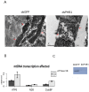
Figure 5. ArPHB1 knockdown resulted in aberrant mitochondrial function.
A, mitochondrial morphology by TEM (magnification, 24000×; scale bar = 0.5 µm). Mitochondria are denoted by red arrows. B, Real-time PCR of mitochondrial genes (ATP6, ATP synthase F0 subunit 6; ND5, NADH dehydrogenase 5; and Cyto B, cytochrome b). Results were normalized to α-tubulin. Means ± SD are plotted. P<0.05. The primers used for real-time PCR are listed in Table S1. C, Western blot analysis of ATPase β subunit.
Figure 6
Figure 6. Involvement of PHB1 in the degradation of yolk platelets.
A, SDS-PAGE analysis of isolated yolk platelet lysates. Proteins were stained by Coomassie brilliant blue R-250. 0 h, 0 h after incubation; 6 h, 6 h after incubation; 12 h, 12 h after incubation; 24 h, 24 h after incubation. B, Yolk platelets were isolated by density centrifugation and probed with an anti-PHB1 antibody. An anti-VGN antibody was used as the control. 0 h, 0 h after incubation of cysts; 6 h, 6 h after incubation of cysts; 12 h, 12 h after incubation of cysts; 24 h, 24 h after incubation of nauplii. C. Yolk protein degradation during embryonic development. 0 h, 0 h after incubation of cysts; 6 h, 6 h after incubation of cysts; 12 h, 12 h after incubation of cysts; 24 h, 24 h after incubation D, Western blot analysis of yolk protein degradation in PHB1 knockdown nauplii. E, TEM analysis showed aberrant yolk platelet degradation in the PHB1 knockdown group (magnification 1250×, scale bar = 1 µm). F, Immunoprecipitation with an anti-PHB1 antibody using isolated yolk platelet lysates (12 h after incubation of cysts). The precipitates were then probed with an anti-ubiquitin antibody.

References

    1. Back JW, Sanz MA, De Jong L, De Koning LJ, Nijtmans LGJ, et al. (2002) A structure for the yeast prohibitin complex: Structure prediction and evidence from chemical crosslinking and mass spectrometry. Protein Science 11: 2471–2478. - PMC - PubMed
    1. Mishra S, Murphy LC, Nyomba BLG, Murphy LJ (2005) Prohibitin: a potential target for new therapeutics. Trends in Molecular Medicine 11: 192–197. - PubMed
    1. Faller DV, Zhang BH, Sheng W (2002) Prohibitin recruits the chromatin-remodeling ATPases Brg-1 and Brm for repression of E2F-driven transcription and cell proliferation. Blood 100: 738A–738A.
    1. Fusaro G, Wang S, Chellappan S (2002) Differential regulation of Rb family proteins and prohibitin during camptothecin-induced apoptosis. Oncogene 21: 4539–4548. - PubMed
    1. Wang S, Nath N, Fusaro G, Chellappan S (1999) Rb and prohibitin target distinct regions of E2F1 for repression and respond to different upstream signals. Molecular and Cellular Biology 19: 7447–7460. - PMC - PubMed

Publication types

LinkOut - more resources