Review
. 2014 Dec:29:178-86.
doi: 10.1016/j.conb.2014.09.010. Epub 2014 Oct 10.

Neural control of brain state

Affiliations
Review

Neural control of brain state

Edward Zagha et al. Curr Opin Neurobiol. 2014 Dec.

Abstract

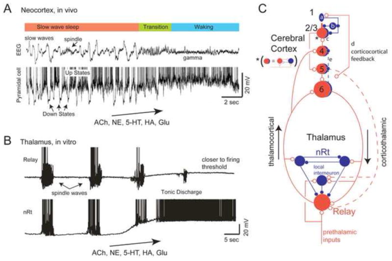
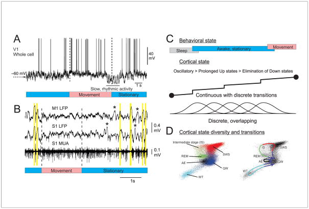
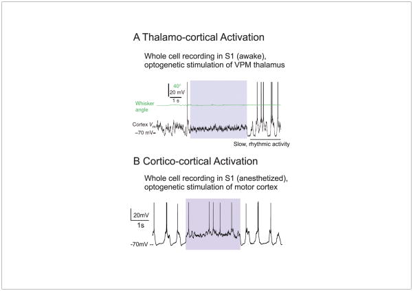
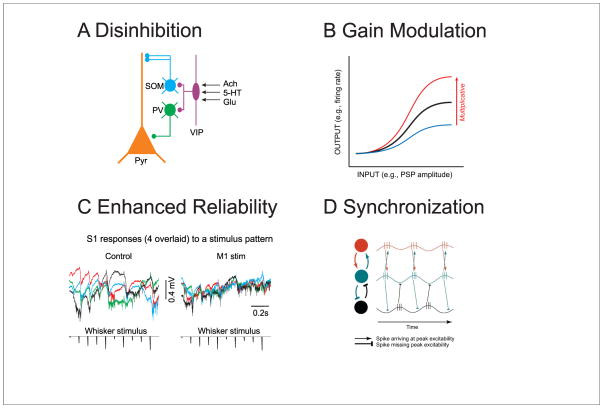
How the brain takes in information, makes a decision, and acts on this decision is strongly influenced by the ongoing and constant fluctuations of state. Understanding the nature of these brain states and how they are controlled is critical to making sense of how the nervous system operates, both normally and abnormally. While broadly projecting neuromodulatory systems acting through metabotropic pathways have long been appreciated to be critical for determining brain state, more recent investigations have revealed a prominent role for fast acting neurotransmitter pathways for temporally and spatially precise control of neural processing. Corticocortical and thalamocortical glutamatergic projections can rapidly and precisely control brain state by changing both the nature of ongoing activity and by controlling the gain and precision of neural responses.


Figures

Figure 1
Figure 1
Cortical and thalamocortical networks exhibit state-dependent changes in network activity. A. During slow wave sleep, the EEG and local cortical field potential is dominated by slow waves, which represent the occurrence of Up and Down states in the local network. The transition to waking is associated with the abolition of the Down states, and the enhancement of higher frequency rhythms such as gamma waves. Several neurotransmitters have been implicated in this transition including acetylcholine (ACh), norepinephrine (NE), serotonin (5-HT), histamine (HA), and glutamate (Glu). Illustrated is the local field potential and intracellular recording from a pyramidal cell during the transition from slow wave sleep to waking. B. Thalamic circuits generate sleep spindle waves as a reverberant interaction of the glutamatergic relay cells and the GABAergic inhibitory neurons of the thalamic reticular nucleus (nRt). The combined action of several neurotransmitters, including ACh, NE, 5-HT, HA, and Glu, can depolarize thalamic circuits out of the sleep-like mode into a state of tonic discharge or ready to discharge. One major mechanism of this depolarization is the reduction of K+ conductances that are active at rest. C. Schematic diagram illustrating major intracortical, intrathalamic, and corticothalamic pathways. Neuromodulatory transmitter systems contact all of these elements and can modulate each in unique ways. A common motif in the cortex is the reciprocal connections of excitatory (red neurons) and inhibitory (blue neurons) neurons (indicated by the asterisk). Recent investigations [–57,82] reveal that VIP interneurons (a) in or near layer 1 can inhibit somatostatin (b) and parvalbumin (c) containing interneurons, resulting in disinhibition of pyramidal cells. Corticocortical connections (d) may specifically engage this disinhibitory circuit. Interlaminar projections within the cortex are not only excitatory, but can also be inhibitory (e), and the activation of this pathway can result in gain modulation [32]. A is from [1]; B is from [50].
Figure 2
Figure 2
Characterizing state changes in behaving mice. A. Whole cell recordings from a pyramidal cell in the primary visual cortex of an awake mouse reveal that movement (walking) is associated with a depolarization of the membrane potential and a suppression of low frequency fluctuations. B. Simultaneous local field potential and multiple unit recordings from primary motor and somatosensory cortex of a mouse in the transition from stationary quiescence to movement (whisking). During quiescence, the cortex exhibits synchronized off periods reminiscent of Down states (yellow bars). These putative Down states may occur locally (e.g. asterisks). Whisking is associated with a suppression of these silent periods and the tonic activation of cortical circuits. C. Behavioral and cortical states are often viewed as exhibiting continuous changes delineated by abrupt transitions, although there may also exist multiple overlapping, yet discrete, states and substates. D. Characterization of behavioral state in rodents by principle component analysis of the activity of multiple brain areas reveals the major sleep-waking states seen behaviorally. Note that although the states exist within their own portions of state-space, they are not completely distinct and separate (left). Movement between states follows repeated paths (right). Abbreviations: AE: active exploration; IS: intermediate stage; REM: rapid eye movement sleep; SWS: slow wave sleep; QW: quiet wake; WT: whisker twitching. A from [9]; B unpublished data (EZ, DM); D from [23].
Figure 3
Figure 3
The stimulation of glutamatergic pathways can result in the rapid activation of cortical networks. A. Whole cell recording from a cortical pyramidal cell during the optogenetic stimulation of thalamus (colored box). During thalamic stimulation, the cortical neuron is rapidly and tonically depolarized and slow fluctuations are suppressed. B. A similar effect is observed upon stimulation of feedback projections from primary motor cortex (M1) to primary somatosensory cortex (S1). Note that both responses exhibit rapid onset and offset kinetics and result in changes in cortical network activity that is similar to arousal, movement, and attention. A is from [14]; B is from [8].
Figure 4
Figure 4
Circuit effects of rapid and slow modulation of cortical state. A. Recent investigations have revealed that disinhibition may be a significant mechanism modulating the responsiveness of cortical pyramidal cells [–57,82]. The proposed microcircuit consists of VIP-containing interneurons inhibiting SOM and PV interneurons, resulting in enhanced responsiveness of postsynaptic pyramidal cells. VIP interneurons are modulated by several ionotropic pathways (nicotinic, 5HT3A, glu), which may allow for the rapid modulation of these neurons. B. Multiplicative gain modulation is a major mechanism by which the input-output relationship of cortical neurons may be modulated. Multiplicative gain modulation can be achieved by changes in the mean membrane potential in the presence of membrane potential variance [61]. C. Suppression of ongoing fluctuations in network activity can result in a significant increase in the reliability of cortical responses to sensory stimuli. Illustrated here are the local field potentials evoked in S1 in response to whisker stimulation either with (M1 stim) or without (control) optogenetic stimulation of feedback pathways from M1 to S1. D. The activation of cortical networks may result in selective propagation of neuronal activity by enhancing synchronization, which allows temporal summation of synaptic responses to bring the postsynaptic neuron to firing threshold. B is adapted from [43]; C is from [8]; D is from [75].

References

    1. Steriade M, Timofeev I, Grenier F. Natural waking and sleep states: a view from inside neocortical neurons. J Neurophysiol. 2001;85:1969–1985. - PubMed
    1. Beloozerova IN, Sirota MG, Swadlow HA. Activity of different classes of neurons of the motor cortex during locomotion. J Neurosci. 2003;23:1087–1097. - PMC - PubMed
    1. O’Connor DH, Peron SP, Huber D, Svoboda K. Neural activity in barrel cortex underlying vibrissa-based object localization in mice. Neuron. 2010;67:1048–1061. - PubMed
    1. Sakata S, Harris KD. Laminar-dependent effects of cortical state on auditory cortical spontaneous activity. Front Neural Circuits. 2012;6:109. - PMC - PubMed
    1. Crochet S, Petersen CC. Correlating whisker behavior with membrane potential in barrel cortex of awake mice. Nat Neurosci. 2006;9:608–610. - PubMed

Publication types