. 2014 Oct 13;9(10):e107551.
doi: 10.1371/journal.pone.0107551. eCollection 2014.

Angiogenic potential of vitreous from Proliferative Diabetic Retinopathy and Eales' Disease patients

Affiliations

Angiogenic potential of vitreous from Proliferative Diabetic Retinopathy and Eales' Disease patients

Ponnalagu Murugeswari et al. PLoS One. .

Abstract

Purpose: Proliferative Diabetic Retinopathy (PDR) and Eales' Disease (ED) have different aetiologies although they share certain common clinical symptoms including pre-retinal neovascularization. Since there is a need to understand if the shared end-stage angiogenic pathology of PDR and ED is driven by common stimulating factors, we have studied the cytokines contained in vitreous from both patient groups and analyzed the angiogenic potential of these samples in vitro.

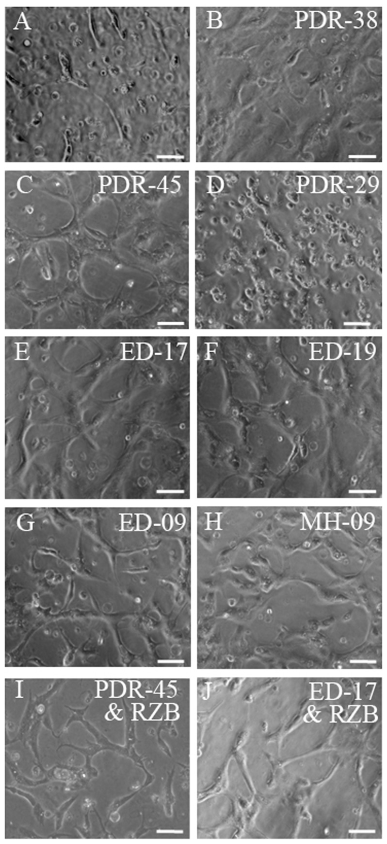
Material and methods: Vitreous samples from patients with PDR (n = 13) and ED (n = 5) were quantified for various cytokines using a cytokine biochip array and sandwich ELISA. An additional group of patients (n = 5) with macular hole (MH) was also studied for comparison. To determine the angiogenic potential of these vitreous samples, they were analyzed for their ability to induce tubulogenesis in human microvascular endothelial cells. Further, the effect of anti-VEGF (Ranibizumab) and anti-IL-6 antibodies were studied on vitreous-mediated vascular tube formation.

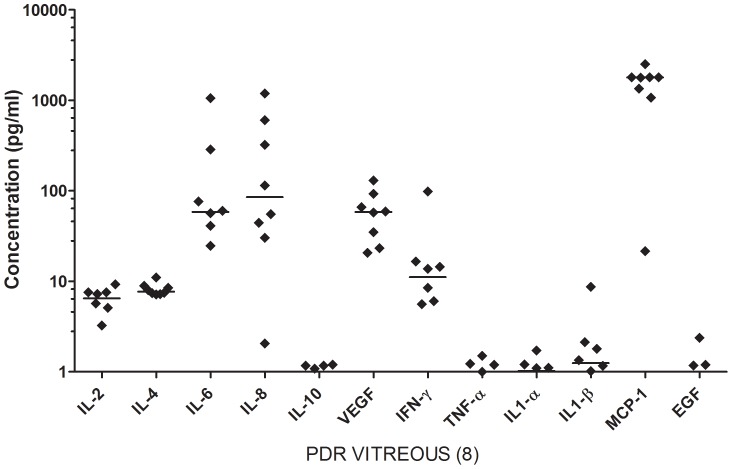
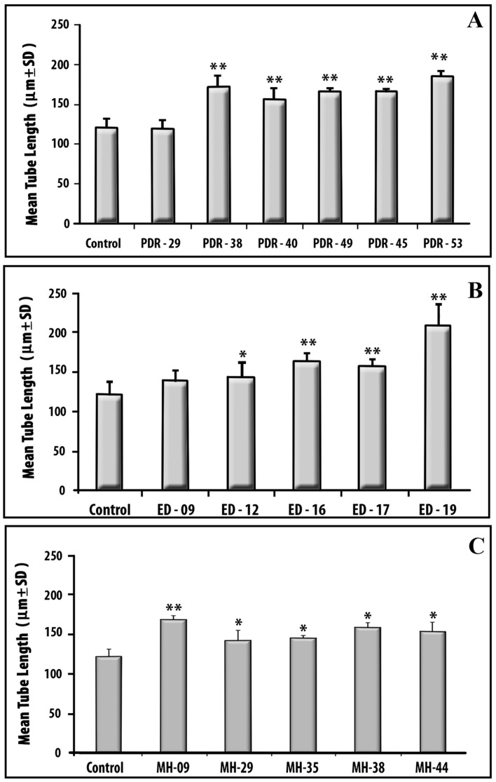
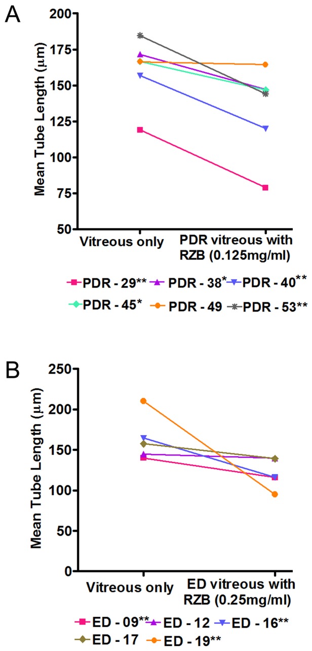
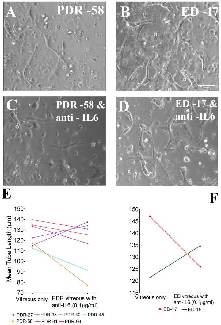
Results: Elevated levels of IL-6, IL-8, MCP-1 and VEGF were observed in vitreous of both PDR and ED when compared to MH. PDR and ED vitreous induced greater levels of endothelial cell tube formation compared to controls without vitreous (P<0.05). When VEGF in vitreous was neutralized by clinically-relevant concentrations of Ranibizumab, tube length was reduced significantly in 5 of 6 PDR and 3 of 5 ED samples. Moreover, when treated with IL-6 neutralizing antibody, apparent reduction (71.4%) was observed in PDR vitreous samples.

Conclusions: We have demonstrated that vitreous specimens from PDR and ED patients share common elevations of pro-inflammatory and pro-angiogenic cytokines. This suggests that common cytokine profiles link these two conditions.


Conflict of interest statement

Competing Interests: Co-author Alan Stitt is a PLOS ONE Editorial Board member. This does not alter the authors' adherence to PLOS ONE Editorial policies and criteria.

Figures

Figure 1
Figure 1. Scatter plot showing the distribution levels of 12 cytokines in vitreous from PDR (n = 8) patients, quantified using cytokine bio-chip array.
Each sample represents the mean of duplicates. Solid line indicates the median. PDR-Proliferative diabetic retinopathy; ED- Eales' disease. IL - Interleukins, VEGF- Vascular endothelial growth factor, IFN-γ- Interferon gamma, TNF-α - Tumour necrosis factor alpha, MCP-1 - Monocyte chemoattractant protein-1, EGF - Epidermal growth factor.
Figure 2
Figure 2. Representative phase images of tube formation induced by vitreous from PDR/ED/MH patients in human dermal microvascular endothelial cell (HMEC).
2×10⁵ HMECs in triplicate were exposed to vitreous alone or with RZB (0.125 mg/ml). Tube formation was observed and images were captured after 48 hours incubation. Each panel shows a part of the representative well. The tube length was quantified by NIS-Elements software (Nikon). Scale bar  = 100 µm. A. Control (without vitreous); B and C - PDR vitreous-induced tube formation, which had high levels of VEGF/IL-6/MCP-1; D-PDR vitreous with trace levels of cytokines showing a very few tube formation. E- G - ED vitreous-induced tube formation as in PDR. H - MH vitreous. I and J are images of vascular tubes in the presence of PDR/ED vitreous and anti-VEGF antibody, showing reduction in tube length compared to C and E respectively. Number in the images denotes the patient ID as in table 1. PDR - Proliferative diabetic retinopathy; ED- Eales' disease; MH- Macular Hole. RZB – Ranibizumab.
Figure 3
Figure 3. Angiogenic potential of vitreous in capillary tube formation.
Experimental details are as in Fig. 2. (A) PDR vitreous, (B) ED vitreous, (C) MH vitreous. Bar graph shows the mean concentration of the triplicate of each sample. PDR-proliferative diabetic retinopathy; ED- Eales' disease; MH- Macular Hole. Number in X-axis denotes the patient number as in table 1. **P<0.001; *P<0.05.
Figure 4
Figure 4. Effect of anti-VEGF antibody on vitreous-induced tube formation.
HMECs were mixed with PDR vitreous in presence of RZB (in triplicate cultures). Tube formation was observed and images were captured after 48 hours incubation. (A) 0.125 mg/ml of RZB with PDR vitreous (B) 0.25 mg/ml of RZB with ED vitreous. The tube length was quantified by Nikon NIS-Elements software. Number in the legend denotes the patient ID as in table 1. PDR - Proliferative diabetic retinopathy; ED- Eales' disease; RZB-Ranibizumab. **P<0.001;*P<0.05.
Figure 5
Figure 5. Effects of anti-IL-6 on vitreous-induced tube formation.
HMECs were mixed with PDR or ED vitreous with or without anti-IL6 (0.1 µg/ml). Tube formation was observed and images were captured after 48 hours of incubation (in duplicate cultures). A and B are PDR or ED vitreous-induced tube formation. C and D are images of vascular tubes of corresponding vitreous with anti-IL6. E and F – shows the effect of anti-IL6 on vascular tube length for 6 PDR (E) and 2 ED (F) vitreous samples. The tube length was quantified by NIS-Elements software (Nikon). Number in the image denotes the patient ID as in table 1. PDR-Proliferative diabetic retinopathy; ED- Eales' disease. Scale bar  = 100 µm.

References

    1. Das T, Biswas J, Kumar A, Nagpal PN, Namperumalsamy P, et al. (1994) Eales' disease. Indian J Ophthalmol 42: 3–18. - PubMed
    1. Murthy KR, Abraham C, Baig SM,Badrinath SS (1977) Eales' disease. Proc All Ind Ophthalmol. pp. 323.
    1. Atmaca LS, Batioglu F, Atmaca Sonmez P (2002) A long-term follow-up of Eales' disease. Ocul Immunol Inflamm 10: 213–221. - PubMed
    1. Murugeswari P, Shukla D, Rajendran A, Kim R, Namperumalsamy P, et al. (2008) Proinflammatory cytokines and angiogenic and anti-angiogenic factors in vitreous of patients with proliferative diabetic retinopathy and eales' disease. Retina 28: 817–824. - PubMed
    1. Ferrara N, Hillan KJ, Gerber HP, Novotny W (2004) Discovery and development of bevacizumab, an anti-VEGF antibody for treating cancer. Nat Rev Drug Discov 3: 391–400. - PubMed

Publication types

Supplementary concepts