. 2014 Oct 23;9(2):581-90.
doi: 10.1016/j.celrep.2014.09.013. Epub 2014 Oct 9.

Development of the fetal bone marrow niche and regulation of HSC quiescence and homing ability by emerging osteolineage cells

Affiliations

Development of the fetal bone marrow niche and regulation of HSC quiescence and homing ability by emerging osteolineage cells

Süleyman Coşkun et al. Cell Rep. .

Abstract

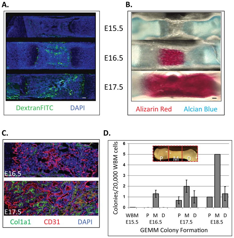
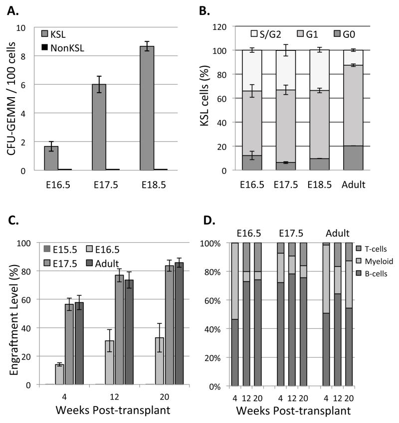
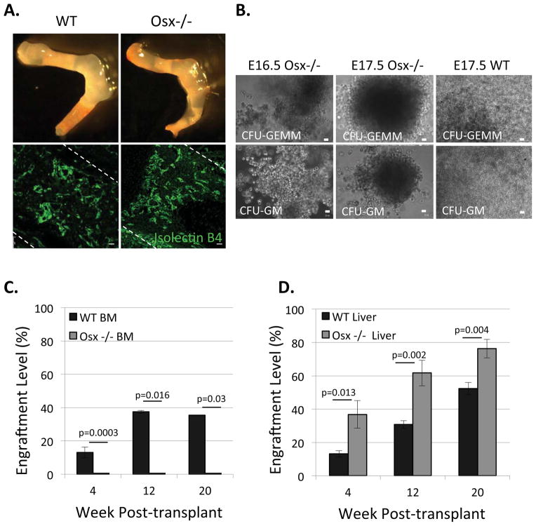
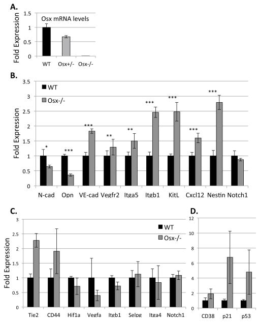
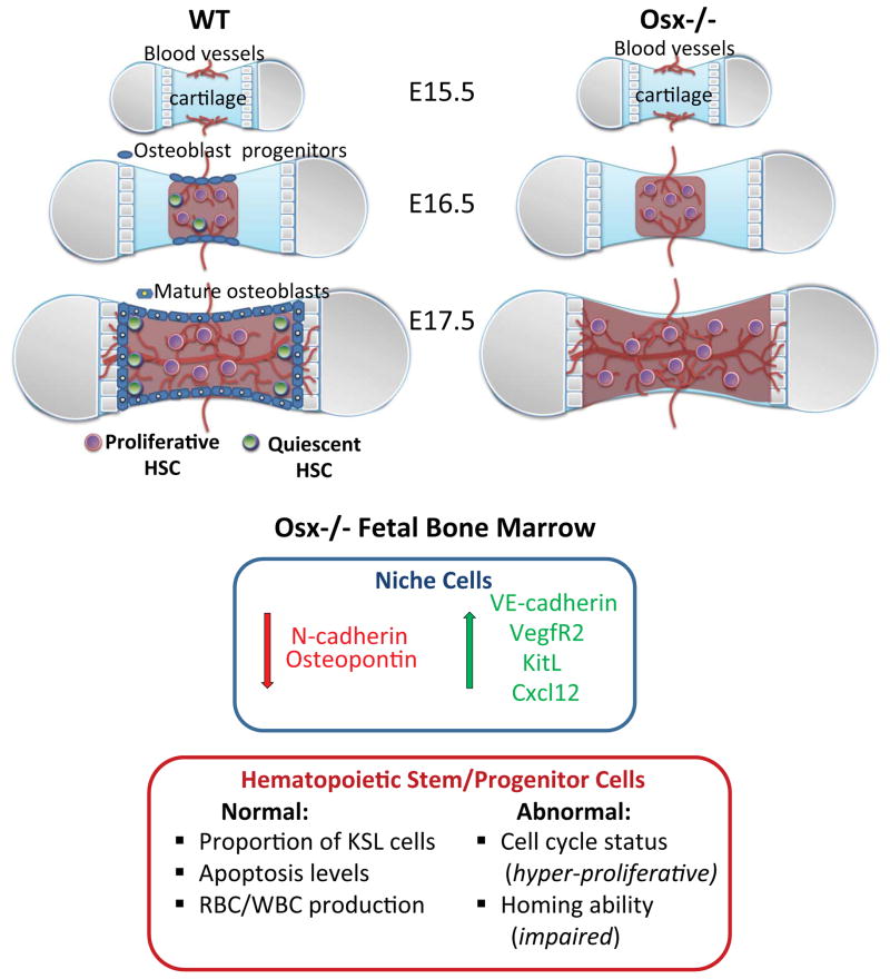
Hematopoietic stem cells (HSCs) reside within a specialized niche where interactions with vasculature, osteoblasts, and stromal components regulate their self-renewal and differentiation. Little is known about bone marrow niche formation or the role of its cellular components in HSC development; therefore, we established the timing of murine fetal long bone vascularization and ossification relative to the onset of HSC activity. Adult-repopulating HSCs emerged at embryonic day 16.5 (E16.5), coincident with marrow vascularization, and were contained within the c-Kit(+)Sca-1(+)Lin(-) (KSL) population. We used Osterix-null (Osx(-/-)) mice that form vascularized marrow but lack osteolineage cells to dissect the role(s) of these cellular components in HSC development. Osx(-/-) fetal bone marrow cells formed multilineage colonies in vitro but were hyperproliferative and failed to home to and/or engraft transplant recipients. Thus, in developing bone marrow, the vasculature can sustain multilineage progenitors, but interactions with osteolineage cells are needed to regulate long-term HSC proliferation and potential.


Figures

Figure 1
Figure 1. Fetal bone is vascularized within the middle region at E16.5 where hematopoietic stem/progenitor cell (HSPC) activity is initially detected
A) Mouse E15.5–17.5 fetuses were injected with 100 μL Dextran-FITC (70 kDa) via superficial temporal vein to reveal functional blood vessels. Fetal femurs were avascular at E15.5, but perfused by E16.5, specifically in the middle regions (Longitudional bone cross section, Green: Dextran-FITC, Blue: nuclear DAPI. Bar = 100 μm). B) Composition of fetal bone was revealed via alizarin red (mineralized tissue) and alcian blue (cartilage) staining. Calcification was apparent by E16.5 in the middle regions, and present throughout the tissue by E17.5. C) At E16.5, Col1a1-positive mature osteoblasts were detected at the periosteum, and throughout the bone at E17.5 (Bar = 100 μm). D) HSPC activity was first detected at E16.5 in the middle regions of bone. Multi-lineage colonies containing granulocyte, erythrocyte, monocyte, megakaryocyte (CFU-GEMM) were scored from 20,000 cells/well for each embryonic time points by Methocult® assay (Data represent mean ± SEM, N=3).
Figure 2
Figure 2. Fetal bone marrow hematopoietic activity is restricted to the KSL population, which is more actively proliferating than adult bone marrow KSL and gives rise to all blood lineages
A) 100 KSL and 1000 NonKSL cells from fetal bone marrow were sorted and cultured in Methocult®; only KSL cells exhibited multi-lineage colony forming activity (Data represent mean ± SEM, N=3; p=0.007 (E16.5), p=0.0005 (E17.5) and p=0.00001 (E18.5)). B) Cell cycle distribution of the fetal and adult bone marrow KSL cells was revealed using PyroninY/Hoechst dyes and flow cytometry. KSL cells were more actively proliferating in fetal vs. adult bone marrow (Data represent mean ± SEM, N=3). C) Engraftment levels were determined at 4, 12 and 20 weeks post-transplantation by analyzing peripheral blood CD45.2 (donor) and CD45.1 (recipient) levels. LT-HSC were first present at E16.5 and showed engraftment levels up to 35% at 20 week post-transplant. By E17.5, engraftment level was comparable to adult bone marrow HSC. (Data represent mean ± SEM, N=3; Differences between E16.5 and either E17.5 or adult were significant; p=0.00085 (week 4), p=0.00326 (week 12) and p=0.00503 (week 20). D) Lineage analysis revealed fetal bone marrow cells (E16.5–17.5) gave rise to all blood lineages in vivo. (Data represent mean ± SEM, N=3).
Figure 3
Figure 3. Osx−/− fetal bone marrow cells form multi-lineage CFU-GEMM colonies in vitro, but fail to repopulate transplanted recipients
A) Osx−/− fetal long bones lack osteoblasts and have structural defects compared to wild type (WT), however, they have a normally vascularized marrow, as revealed by isolectin B4-FITC staining. Bar=20 μm B) Representative images of CFU-GEMM and CFU-GM colonies formed from E16.5 and E17.5 Osx−/− fetal bone marrow cells, similar to E17.5 WT fetal bone marrow cells. C) 400,000 WT and Osx−/− WBM cells at E17.5 were transplanted into sublethally irradiated neonates and engraftment levels were determined by analyzing recipients’ peripheral blood up to 20 week post-transplant; E17.5 Osx−/− WBM cells did not engraft. (Data represent mean ± SEM, N=3; p=0.0003 (week 4), p=0.016 (week12) and p=0.03 (week 20). D) WT and Osx−/− derived E17.5 fetal liver cells repopulated transplant recipients’ peripheral blood in vivo (Data represent mean ± SEM, N=3; p=0.013 (week 4), p=0.002 (week12) and p=0.004 (week 20).
Figure 4
Figure 4. Fetal Bone Marrow Cells Derived from Osx−/− Mutants Exhibit a Homing Defect
A) P0-P2 recipient neonates were euthanized 24 h post-transplantation and bone marrow and liver cells were analyzed with flow cytometry for donor/recipient cell ratios. B) Fetal liver cells from E17.5 WT and Osx−/− mutants exhibited similar homing to liver and bone marrow of WT recipients. C) In contrast, fetal WBM cells derived from E17.5 Osx−/− mutants showed significantly reduced homing compared to WT (Data represent mean ± SEM, N=3).
Figure 5
Figure 5. Gene expression studies revealed that genes specific to the bone marrow niche and HSPC are dysregulated in osterix mutants
A) Gene expression analysis (via qPCR) was performed on E17.5 WT and Osx−/− WBM cells. Osx expression was ~40% decreased in Osx+/− fetuses and absent in Osx−/− mutants. B) N-cadherin (cdh2) and osteopontin (opn) were significantly downregulated in Osx−/− mutants; whereas, Cxcl12, kitL (SCF), nestin, vascular endothelial cell adhesion protein, VE-cadherin (cdh5), vascular endothelial growth factor receptor 2 (vegfr2), integrin subunits β1 (itgb1) and α5 (itga5) were significantly upregulated compared to WT (Asterix (*), (**), and (***) denotes p≤0.01, p≤0.001 and p≤0.0001 respectively). β-actin gene expression was used as an internal control. For each gene of interest, triplicates from four different cDNA samples were analyzed (Data represent mean ± SEM, N=4, triplicates for each sample). C–D) KSL cells were isolated from the hindlimbs of E17.5 WT and Osx−/− mutants and analyzed in 10-cell pools. In Osx−/− KSL cells, the expression of CD38, p21, p53, as well as Tie2 and CD44, trended toward higher than wild type littermates; whereas, expression of Hif1α and VegfA trended toward lower. β-actin served as an internal control. Statistical analyses were done using the two-tailed unpaired student t-test (Data represent mean ± SEM, N=4; p=0.30 (CD38), p=0.15 (p21), p=0.30 (p53), p=0.17 (Tie-2), p=0.28 (CD44), p=0.44 (Hif1a), p=0.33 (Vegfa), p=0.5 (itgb1), p=0.79 (Selpg), p=0.42 (itga4), p=0.29 (notch1).
Figure 6
Figure 6. Working model
In wild type (WT) fetal long bones (femur), adult repopulating HSC are first present at E16.5, coincident with vascular perfusion, localized to vascularized regions of bone, and are highly proliferative relative to adult HSC. Osteoblast progenitors are also present within fetal bone at this stage, but appear to be restricted to the periosteum. From E17.5 onward, HSC are also detected within the proximal and distal ends of bone that are fully vascularized and contain mature osteoblasts that are thought to regulate HSC quiescence. In Osx−/− fetal bone marrow, osteoblasts and osteolineage cells do not develop, and there is increased expression of vascular niche associated genes, including VE-cadherin, Vegfr2, KitL and cxcl12, as well as decreased expression of N-cadherin and osteopontin, which suggests increased representation of vascular endothelial cells in this microenvironment in osterix-null mutants. HSPC phenotype and function are also altered in the osteolineage cell-deficient microenvironment. Although KSL cells are present in equal numbers compared to WT, and exhibit similar apoptosis levels, KSL cells isolated from Osx−/− fetal bone marrow exhibited dysregulated cell cycle progression and defective homing ability. These data suggest that osteolineage cells in fetal bone marrow play a critical role in establishing and sustaining LT-HSC phenotype and function during development.

References

    1. Arai F, Hirao A, Ohmura M, Sato H, Matsuoka S, Takubo K, Ito K, Koh GY, Suda T. Tie2/angiopoietin-1 signaling regulates hematopoietic stem cell quiescence in the bone marrow niche. Cell. 2004;118:149–161. - PubMed
    1. Bowie MB, McKnight KD, Kent DG, McCaffrey L, Hoodless PA, Eaves CJ. Hematopoietic stem cells proliferate until after birth and show a reversible phase-specific engraftment defect. J Clin Invest. 2006;116:2808–2816. - PMC - PubMed
    1. Calvi LM, Adams GB, Weibrecht KW, Weber JM, Olson DP, Knight MC, Martin RP, Schipani E, Divieti P, Bringhurst FR, et al. Osteoblastic cells regulate the haematopoietic stem cell niche. Nature. 2003;425:841–846. - PubMed
    1. Challen GA, Boles N, Lin KK, Goodell MA. Mouse hematopoietic stem cell identification and analysis. Cytometry A. 2009;75:14–24. - PMC - PubMed
    1. Chen J, Shi Y, Regan J, Karuppaiah K, Ornitz DM, Long F. Osx-Cre targets multiple cell types besides osteoblast lineage in postnatal mice. PLoS One. 2014;9:e85161. - PMC - PubMed

Publication types

MeSH terms