. 2015 Aug;41(5):613-30.
doi: 10.1111/nan.12189. Epub 2015 Apr 23.

Identification of clinical target areas in the brainstem of prion-infected mice

Affiliations

Identification of clinical target areas in the brainstem of prion-infected mice

Ilaria Mirabile et al. Neuropathol Appl Neurobiol. 2015 Aug.

Abstract

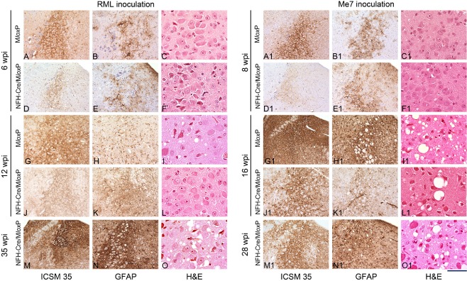
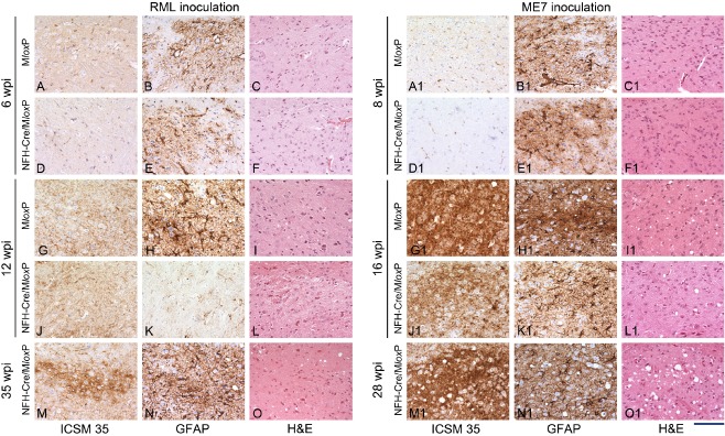
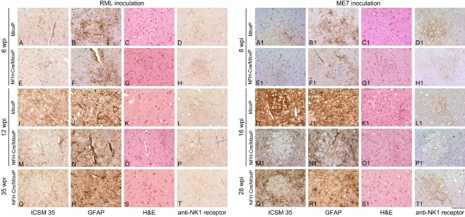
Aims: While prion infection ultimately involves the entire brain, it has long been thought that the abrupt clinical onset and rapid neurological decline in laboratory rodents relates to involvement of specific critical neuroanatomical target areas. The severity and type of clinical signs, together with the rapid progression, suggest the brainstem as a candidate location for such critical areas. In this study we aimed to correlate prion pathology with clinical phenotype in order to identify clinical target areas.

Method: We conducted a comprehensive survey of brainstem pathology in mice infected with two distinct prion strains, which produce different patterns of pathology, in mice overexpressing prion protein (with accelerated clinical onset) and in mice in which neuronal expression was reduced by gene targeting (which greatly delays clinical onset).

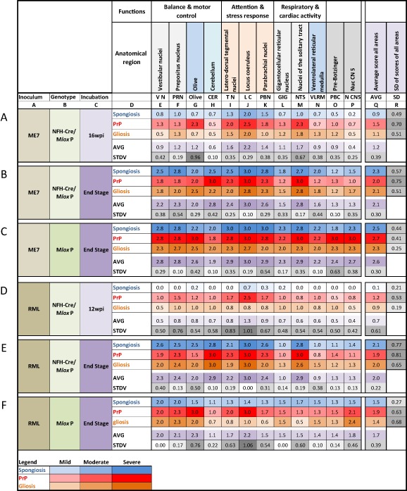
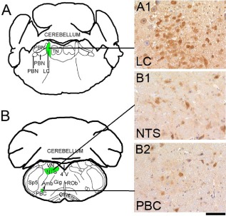
Results: We identified specific brainstem areas that are affected by prion pathology during the progression of the disease. In the early phase of disease the locus coeruleus, the nucleus of the solitary tract, and the pre-Bötzinger complex were affected by prion protein deposition. This was followed by involvement of the motor and autonomic centres of the brainstem.

Conclusions: Neurodegeneration in the locus coeruleus, the nucleus of the solitary tract and the pre-Bötzinger complex predominated and corresponded to the manifestation of the clinical phenotype. Because of their fundamental role in controlling autonomic function and the overlap with clinical signs in sporadic Creutzfeldt-Jakob disease, we suggest that these nuclei represent key clinical target areas in prion diseases.

Keywords: brainstem; clinical target areas; cre-lox system; locus coeruleus; neurodegeneration; prions.


Figures

Figure 1
Figure 1
Abnormal PrP accumulation in the locus coeruleus of RML‐ and ME7‐inoculated MloxP and NFHCre/MloxP mice. Early prion pathology in the brainstem of RML and ME7 prion‐inoculated MloxP (A–D) and NFHCre/MloxP (E,F) mice, time‐culled at an early asymptomatic stage (RML, 6 wpi; ME7, 8 wpi), manifested with deposition of disease‐associated prion protein (red = intermediate, pink = mild, red outline = very mild) and reactive gliosis [dark yellow = intermediate (A); light yellow = mild (B,E,F)] in the locus coeruleus (LC), the nucleus of the solitary tract (NTS) and the pre‐Bötzinger complex (PBC). No spongiform changes were observed at this time point. (A,B,E,F) Schematic representations adapted from the Paxinos Mouse Atlas, −5.34 mm (A,E) and −6.84 (B,F) from Bregma. (C,D) Synaptic PrP deposition in the LC, in RML‐inoculated MloxP mice (A), and granular deposits in ME7‐inoculated MloxP mice (D). 4 V, fourth ventricle; Amb, nucleus ambiguous; LC, locus coeruleus; NTS, nucleus of the solitary tract; PBC, pre‐Bötzinger complex; PBN, parabrachial nuclei; ROb, raphe obscurus; Sp5, spinal nucleus of the V nerve; TN, tegmental nuclei; VN, vestibular nuclei. Scale bar below D = 160 μm.
Figure 2
Figure 2
Survival of RML‐ and ME7‐ inoculated MloxP and NFHCre/MloxP mice. Mice were inoculated with RML or ME7 prions at 1 week of age. MloxP mice (blue line) became terminally ill at approximately 12 wpi (RML, A) or at approximately 17 wpi (ME7, B). In contrast to previous experiments, recombined NFH‐Cre/MloxP mice (red) ultimately developed prion disease at approximately 35 weeks (A) or approximately 29 weeks (B) but both showed significant delay of their incubation time (P < 0.001).
Figure 3
Figure 3
Progression of prion pathology in RML‐ and ME7‐inoculated MloxP and NFHCre/MloxP mice. Schematic drawings of abnormal PrP deposition (pink = mild, red = intermediate, dark red = severe) and spongiosis (pale blue = mild, blue = intermediate, dark blue = severe) of the brainstem nuclei analysed, at four representative levels from coordinates −5.34 mm to −7.64 mm caudal from bregma. The inoculum is shown on the left margin. Genotype and incubation times on the right margin. (A–D) In terminally ill RML‐inoculated MloxP mice (12 wpi) PrP deposition is seen in different areas of the brainstem, including the locus coeruleus and the olive; prepositus nuclei and tegmental nuclei; and pre‐Bötzinger complex; spongiosis corresponds to PrP deposition in most areas. (E–H) In terminally ill ME7 inoculated MloxP mice (16 wpi), abnormal PrP deposition was severe in all the nuclei examined, except the gigantocellular nucleus, the ventral medulla and the cerebellum, where it was intermediate. Spongiosis pattern corresponds to PrP deposition in most areas. (I–L) RML‐inoculated NFHCre/MloxP mice show a significantly lower overall accumulation at 12 wpi, and spongiosis was nearly absent everywhere, except for the locus coeruleus, where it was mild (pale blue). (M–P) In ME7‐inoculated NFHCre/MloxP mice culled at 16 wpi, abnormal PrP accumulation was slightly stronger than in RML‐inoculated animals and more heterogeneous. Spongiosis was reduced everywhere compared with ME7‐inoculated MloxP mice at end stage of prion disease. Strong PrP deposition and spongiosis was observed in terminally ill RML‐inoculated NFH‐Cre/MloxP mice (35 wpi, Q–T), or in terminally ill ME7‐inoculated NFH‐Cre/MloxP mice (28 wpi) (U –X). 10 N, nucleus of the X nerve; 12 N, nucleus of the XII nerve; 4 V, forth ventricle; Amb, nucleus ambiguous; LC, locus coeruleus; NTS, nucleus of the solitary tract; PBC, pre‐Bötzinger complex; PBN, parabrachial nuclei; PR N, prepositus nucleus; ROb, raphe obscurus; Sp5, Spinal nucleus of the V nerve; TN, tegmental nuclei; VLRM, ventro‐lateral reticular medulla; VN, vestibular nuclei; VN, vestibular nuclei.
Figure 4
Figure 4
Summary scores from multiple individual experiments. The top two rows (unlabelled) show the anatomical brainstem regions and their function. Rows A–F show the experimental groups, by inoculum, genotype and incubation time. Above row A, columns A–R provide reference points for the in text citations of anatomical regions. Each of the rows A–F display the summary scores for spongiosis (blue), PrP deposition (red) and gliosis (orange), averaged from at least three experimental animals. The colour intensity is directly proportional to the average scores and was computed with the ‘conditional formatting’ in MS Excel. Below the individual pathology scores, an average of these values is formed to inform about the overall (average or aggregated) pathology. In addition, the row below this average, the standard deviation is calculated to indicate low or high variation between the three scores. A low score (light grey) indicates that all three pathologies develop synchronously in a given region, whereas a high score (dark grey) indicate that the three pathologies develop asynchronously. On the right, columns Q and R indicate the average and standard deviation across all regions. A low value indicates that all regions show similar pathology across the entire brainstem (homogenous) and a high value indicates that some areas show a stronger deposition than others. The legend on the bottom indicates the colours corresponding to scores mild (value 1), moderate (2) and severe (3).
Figure 5
Figure 5
Progression of prion pathology in the locus coeruleus of RML‐ and ME7‐inoculated MloxP and NFHCre/MloxP mice. Sections were immunostained with ICSM35 antibody for abnormal PrP deposition (left column; RML: A,D,G,J,M; ME7: A1,D1,G1,J1,M1), anti‐GFAP antibody for detection of astrocytosis (middle column; RML: B,E,H,K,N; ME7: B1,E1,H1,K1,N1), and stained with haematoxylin and eosin (H&E) to assess spongiform changes (right column; RML: C,F,I,L,O; ME7: C1,F1,I1,L1,O1). A–O Pathology in RML inoculated brains: at 6 wpi, there is synaptic PrPSc deposition in MloxP mice (A), or a mild granular deposition in depleted NFHCre/MloxP mice (D). No difference in gliosis (B,E) and no spongiosis (C,F). At 12 wpi (G–L), strong accumulation in the LC of terminally ill MloxP mice (G), but not in NFHCre/MloxP mice was seen. Gliosis was stronger in NFHCre/MloxP (K) mice than in control MloxP mice (H). Spongiform changes in MloxP mice (I) but not in NFHCre/MloxP mice (L). M–O terminally ill NFHCre/MloxP mice showed accumulation of PrP, gliosis and spongiosis. A1–O1 Pathology in ME7‐inoculated brains: At 8 wpi the abnormal accumulation in the LC of MloxP mice was intermediate (A1) and less intense in NFHCre‐MloxP mice (D1) and there was mild gliosis and no spongiosis (B1,C1,E1,F1). At 16 wpi, MloxP mice were terminally ill and showed strong PrP accumulation, gliosis and spongiosis in and beyond the LC (G1,H1,I1), whereas asymptomatic NFHCre‐MloxP were less affected (J1,K1,L1). In terminally ill NFHCre‐MloxP mice, there was severe LC pathology (M1,N1,O1). Scale bar = 160 μm in all immunostained sections and 80 μm in the H&E stained sections.
Figure 6
Figure 6
Progression of prion pathology in the nucleus of the solitary tract of RML‐ and ME7‐inoculated MloxP and NFHCre/MloxP mice. Sections were immunostained with ICSM35 antibody for abnormal PrP deposition (left columns) anti‐GFAP antibody for detection of astrocytosis (middle columns) and stained with haematoxylin and eosin (H&E) to assess spongiform changes (right columns). (A–O) RML inoculated MloxP mice (6 wpi) showed mild abnormal PrP accumulation, gliosis and no spongiosis (A–C). At the same time, NFHCre‐MloxP mice showed a similarly mild pathology (D–F). At 12 wpi, terminally ill MloxP mice (G–I) showed more PrP accumulation, strong gliosis and minimal spongiosis. At the same time NFHCre/MloxP mice showed similar PrP accumulation but less gliosis and no spongiosis (J–L). Terminally ill NFHCre‐MloxP mice showed stronger PrP accumulation, gliosis and spongiosis, similar to terminally ill MloxP mice (G–I). A1–O1 ME7‐inoculated mice showed minimal PrP deposition, no spongiosis and moderate gliosis at 8 wpi (A1–F1) and a rapid progression to a more severe pathology at 16 wpi, when MloxP mice are terminal (G1–L1). At end stage (M1–O1), NFHCre/MloxP mice showed severe pathology with abundant PrP, intense gliosis and moderate spongiosis. Scale bar = 160 μm.
Figure 7
Figure 7
Progression of prion pathology in the pre‐Bötzinger complex of RML‐and ME7‐inoculated MloxP and NFHCre/MloxP mice. Sections were immunostained with ICSM35 antibody for abnormal PrP deposition (left column) anti‐GFAP antibody for detection of astrocytosis (second column) and stained with haematoxylin and eosin (H&E) to assess spongiform changes (third column) and NK1 Receptor (right column). A–T RML‐inoculated MloxP mice (6 wpi) showed mild to moderate abnormal PrP accumulation, gliosis and no spongiosis (A–C). At the same time, NFHCre/MloxP mice showed a similarly mild pathology (E–G). At 12 wpi, terminally ill MloxP mice (I–K) showed more PrP accumulation, strong gliosis and minimal spongiosis. At the same time NFHCre/MloxP mice showed similar PrP accumulation but less gliosis and no spongiosis (M–O). Terminally ill NFHCre‐MloxP mice showed still a moderate PrP accumulation, gliosis and spongiosis, similar to terminally ill MloxP mice (I–K). There is some inconsistency of progression, as the gliosis in (E) is stronger than that in (K), indicating a possible variance in transgene recombination efficacy. The right column demonstrates the NK1R immunolabelling of the pre‐Bötzinger complex. A1–T1 ME7‐inoculated MloxP and NFHCre/MloxP mice show minimal PrP deposition, no spongiosis and moderate gliosis at 8 wpi (A1–C1, E1–G1). At 16 wpi more severe pathology, with abundant PrP deposition, severe gliosis and spongiosis was observed in terminally sick MloxP mice (I1K1) but not asymptomatic NFHCre/MloxP mice (M1O1). At end stage (Q1S1), NFHCre/MloxP mice showed severe pathology with abundant PrP, intense gliosis and moderate spongiosis. Right column, NK1R immunolabelling to verify the pre‐Bötzinger complex. Scale bar = 160 μm.
Figure 8
Figure 8
Cre‐mediated recombination in the locus coeruleus (LC), the nucleus of the solitary tract (NTS) and the pre‐Bötzinger complex (PBC) of NFHCre/ROSA26 mice. A,B schematic representations adapted from the Paxinos Mouse Atlas, coordinates −5.34 mm (A) and −6.84 mm (B) from Bregma. A1,B1,B2 β‐galactosidase immunoreactivity in the LC (A1), the NTS (B1) and the PBC (B2) of NFHCre/ROSA26 mice. Immunostaining for β‐galactosidase is characterized by a diffuse accumulation in the cytoplasm as well as few small intracellular cytoplasmic inclusions. In the LC, ca. half of the cells express β‐galactosidase, indicating partial recombination (A1). In the nucleus of the solitary tract (B1) and in the pre‐Bötzinger complex (B2), recombination was less widespread. Scale bar = 80 μm.

References

    1. Bruce ME, McBride PA, Farquhar CF. Precise targeting of the pathology of the sialoglycoprotein, PrP, and vacuolar degeneration in mouse scrapie. Neurosci Lett 1989; 102: 1–6 - PubMed
    1. Bruce ME. Serial studies on the development of cerebral amyloidosis and vacuolar degeneration in murine scrapie. J Comp Pathol 1981; 91: 589–597 - PubMed
    1. Liberski PP, Sikorska B, Hauw JJ, Kopp N, Streichenberger N, Giraud P, Boellaard J, Budka H, Kovacs GG, Ironside J, Brown P. Ultrastructural characteristics (or evaluation) of Creutzfeldt‐Jakob disease and other human transmissible spongiform encephalopathies or prion diseases. Ultrastruct Pathol 2010; 34: 351–361 - PubMed
    1. Kovacs GG, Budka H. Prion diseases: from protein to cell pathology. Am J Pathol 2008; 172: 555–565 - PMC - PubMed
    1. Liberski PP, Ironside JW. An outline of the neuropathology of transmissible spongiform encephalopathies (prion diseases). Folia Neuropathol 2004; 42 (Suppl. B): 39–58 - PubMed

Publication types

LinkOut - more resources