. 2014 Nov 24;34(6):e00154.
doi: 10.1042/BSR20140088.

Apo, Zn2+-bound and Mn2+-bound structures reveal ligand-binding properties of SitA from the pathogen Staphylococcus pseudintermedius

Affiliations

Apo, Zn2+-bound and Mn2+-bound structures reveal ligand-binding properties of SitA from the pathogen Staphylococcus pseudintermedius

Francesca Abate et al. Biosci Rep. .

Abstract

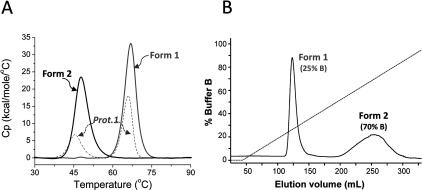
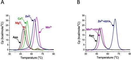
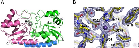
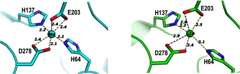
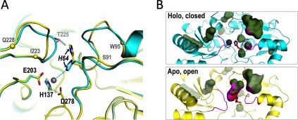
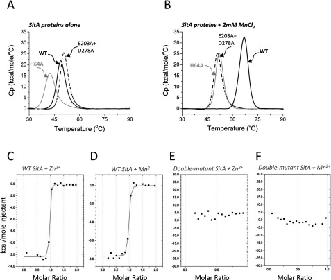
The Gram-positive bacterium Staphylococcus pseudintermedius is a leading cause of canine bacterial pyoderma, resulting in worldwide morbidity in dogs. S. pseudintermedius also causes life-threatening human infections. Furthermore, methicillin-resistant S. pseudintermedius is emerging, resembling the human health threat of methicillin-resistant Staphylococcus aureus. Therefore it is increasingly important to characterize targets for intervention strategies to counteract S. pseudintermedius infections. Here we used biophysical methods, mutagenesis, and X-ray crystallography, to define the ligand-binding properties and structure of SitA, an S. pseudintermedius surface lipoprotein. SitA was strongly and specifically stabilized by Mn2+ and Zn2+ ions. Crystal structures of SitA complexed with Mn2+ and Zn2+ revealed a canonical class III solute-binding protein with the metal cation bound in a cavity between N- and C-terminal lobes. Unexpectedly, one crystal contained both apo- and holo-forms of SitA, revealing a large side-chain reorientation of His64, and associated structural differences accompanying ligand binding. Such conformational changes may regulate fruitful engagement of the cognate ABC (ATP-binding cassette) transporter system (SitBC) required for metal uptake. These results provide the first detailed characterization and mechanistic insights for a potential therapeutic target of the major canine pathogen S. pseudintermedius, and also shed light on homologous structures in related staphylococcal pathogens afflicting humans.


Figures

Figure 1
Figure 1. Differential scanning calorimetry profiles of different grades of SitA purifications
(A) In DSC experiments a Tm value is given by the peak maximum of the scanned melting curve. The DSC profile of the first purified preparation of SitA (‘Prot.1’, dashed grey line), showed that the initial sample generated two peaks (Tm1 45.6°C and Tm2 66.0°C), consistent with two unfolding events, which could potentially be generated either by two different domains within one protein, or by two different forms of the same protein. The latter option was supported by HIC, from which two separate forms of SitA were obtained [Form-1 and Form-2, see panel (B)], each of which generated only one peak in DSC experiments (solid black and grey lines), Tm 47.7°C for Form-2, and Tm 66.9°C for Form-1. (B) The HIC elution profile of SitA, showing clear separation of two forms.
Figure 2
Figure 2. Different binding and reversibility of divalent metal cations with SitA
(A) The purified recombinant apo-SitA protein displays a single peak, Tm=48.7°C (black line), the lowest Tm measured in all these experiments. The DSC data for SitA+2 mM MnCl2 (magenta line, Tm 66.9°C) or SitA+2 mM ZnCl2 (blue line, Tm 64.1°C) displayed a single peak, corresponding to an unfolding event with high Tm. The DSC data for SitA+2 mM CaCl2 (green line, Tm 53.9°C) or 2 mM MgCl2 (red line, 51.2°C) showed much smaller binding-induced increases in Tm. (B) A strong chelating agent (EDTA, 10 mM) was added to SitA previously complexed with 2 mM Mn2+ or 2 mM Zn2+ and the DSC profiles were recorded. While the DSC profile of the Mn2+/EDTA-treated SitA sample (magenta line) revealed an unfolding transition which closely resembled that of metal-free apo-SitA (Tm 48.7°C, black line), the Zn2+/EDTA-treated SitA sample (blue line) revealed a Tm of 70.1°C, which approximately matched the profile of metal-bound SitA. (The reason for the minor difference in Tm between SitA+Zn2+ in the presence and absence of EDTA is currently unclear. Nevertheless, EDTA does not appear to stabilize SitA+Mn2+, revealing that SitA has a different propensity to bind the two metals under these conditions.)
Figure 3
Figure 3. X-ray crystallographic structure of SitA bound to a Mn2+ ion
(A) Cartoon representation of SitA showing the typical class III SBP fold: the N-terminal lobe (green) and C-terminal lobe (pink) are connected by a single long α-helix (blue). The Mn2+ ion, shown as a sphere, binds in a cavity between the two lobes. (B) Details in the ligand-binding pocket of Mn2+-bound SitA, with 2Fo–Fc electron density maps contoured at 2σ (shown as cyan mesh). Bond sticks are coloured by element, with carbon, oxygen, and nitrogen atoms coloured in yellow, red and blue, respectively. The Mn2+ ion is shown as a purple sphere; red spheres show water molecules. Key metal-binding residues are labelled in large italics, close neighbours are labelled in smaller non-italic font.
Figure 4
Figure 4. Atomic details of the SitA-metal cation interactions
The cation-binding sites of Mn2+-bound SitA (cyan, left panel) and Zn2+-bound SitA (green, right panel) reveal that the binding mechanisms are very similar, with a small but notable difference in metal coordination observed for the His64 side chain. Notably, the side chain NE2 atom of His64 is 2.2 Å from the Mn2+ ion, but is shifted >1 Å further away (3.1 Å) from the Zn2+ ion. For each structure, the B-factors of the metal ions refined to values similar to those of the cognate protein atoms involved in metal chelation.
Figure 5
Figure 5. SitA orthologues show high sequence identity and conservation of metal-binding residues
A multiple sequence alignment revealing the high degree of sequence identity (ID) between SitA and its orthologues: SitC in S. epidermidis (68% ID), MntC in S. aureus (65% ID), PsaA in S. pneumoniae (48% ID), MtsA in S. pyogenes (45% ID) and TroA in S. suis (27% ID). The alignments begin at the reactive Cys residue of the lipobox motif conserved in these Gram-positive lipoproteins. Residues are numbered according to full-length SitA, and are shaded black if fully conserved, or are boxed if partially conserved. Metal-chelating residues are indicated with black triangles. Secondary structure elements of SitA (derived from the Mn2+-bound structure) are shown above the sequences. Multiple sequence alignments and figures were prepared using the MAFFT algorithm [59] and ESPript [60].
Figure 6
Figure 6. Differences proximal to the ligand-binding pocket in apo- and holo-SitA
(A) Cartoon representations of the aligned structures of Zn2+-bound SitA (cyan, as in Figure 4) and apo-SitA (yellow) which were observed within the same asymmetric unit. The differences in the metal-binding site of apo- and holo-SitA are most notable in two loops S91–W95 and I223–Q228 (demarcated by Cα spheres on the apo- structure) surrounding the ligand-binding pocket and the outward position of His64 seen in the apo-structure, which is incompatible with the closed holo-structure, since it would clash with Thr225 (pale grey) in the holo-structure. Metal-chelating residues are labelled in black font, with the dynamic His64 in italics. (B) The conformational changes highlighted by the ribbon representations in panel (A) also lead to two rather different surface/cavity landscapes for the apo- and holo-forms of SitA. Here, blob-surfaces show internal cavities and/or surface-exposed tunnels/pockets. Most of the blobs are present in both states, showing that holo-SitA (upper panel) and apo-SitA (lower panel) surfaces/cavities are highly conserved. However, for apo-SitA, the additional large blob in the centre of the lower panel represents the tunnel leading from the apo-SitA surface down into the metal-free binding site. This tunnel disappears upon metal binding and closure, and hence the blob is absent in the upper panel. The flexible loops S91–W95 and I223–Q228 are coloured magenta, while sticks of His64 are coloured red and blue for Cα and nitrogen atoms. The side-chain contributions of the loops and of His64 to lining the walls of the cavities, are shown with magenta and red spots, respectively. All figures were made using Pymol v1.7, using surface ‘cavity’ mode for panel B.
Figure 7
Figure 7. SitA point mutations abolish Mn2+-induced stabilization
(A) DSC experiments performed in the absence of metal ions revealed only a single peak, with similarly low Tm values (40–50°C) for WT (solid black line, Tm 48.7°C) and the SitA mutants H64A (grey line, Tm 42.4) and E203A+D278A (dashed black line, Tm 50.6°C). (B) DSC experiments performed in the presence of 2 mM MnCl2 revealed that the H64A mutant (grey line, Tm 52.0°C) shows a small but notable increase in Tm, whereas the double-mutant E203A+D278A (dashed black line, Tm 51.0°C) shows no change in Tm. The WT protein (solid black line, Tm 66.9°C) exhibits the typical large increase in Tm in presence of Mn2+ ions. (CF) ITC experiments to examine the interaction of wild-type and mutant SitA with Mn2+ and Zn2+ ions. Panels (C) and (D) show the high affinity interaction of Mn2+ and Zn2+ with wild-type SitA; panels (E) and (F) show data for the SitA E203+D278A double-mutant, which presented no detectable interaction with Zn2+ ions or Mn2+ ions.

References

    1. Bannoehr J., Ben Zakour N. L., Waller A. S., Guardabassi L., Thoday K. L., van den Broek A. H., Fitzgerald J. R. Population genetic structure of the Staphylococcus intermedius group: insights into agr diversification and the emergence of methicillin-resistant strains. J. Bacteriol. 2007;189:8685–8692. doi: 10.1128/JB.01150-07. - DOI - PMC - PubMed
    1. Fitzgerald J. R. The Staphylococcus intermedius group of bacterial pathogens: species re-classification, pathogenesis and the emergence of meticillin resistance. Vet. Dermatol. 2009;20:490–495. doi: 10.1111/j.1365-3164.2009.00828.x. - DOI - PubMed
    1. Hill P. B., Lo A., Eden C. A., Huntley S., Morey V., Ramsey S., Richardson C., Smith D. J., Sutton C., Taylor M. D., et al. Survey of the prevalence, diagnosis and treatment of dermatological conditions in small animals in general practice. Vet. Rec. 2006;158:533–539. doi: 10.1136/vr.158.16.533. - DOI - PubMed
    1. Guardabassi L., Schwarz S., Lloyd D. H. Pet animals as reservoirs of antimicrobial-resistant bacteria. J. Antimicrob. Chemother. 2004;54:321–332. doi: 10.1093/jac/dkh332. - DOI - PubMed
    1. Van Hoovels L., Vankeerberghen A., Boel A., Van Vaerenbergh K., De Beenhouwer H. First case of Staphylococcus pseudintermedius infection in a human. J. Clin. Microbiol. 2006;44:4609–4612. doi: 10.1128/JCM.01308-06. - DOI - PMC - PubMed

Publication types

MeSH terms

Associated data

LinkOut - more resources