. 2014 Nov;144(5):357-78.
doi: 10.1085/jgp.201411229. Epub 2014 Oct 13.

Calmodulin enhances ribbon replenishment and shapes filtering of synaptic transmission by cone photoreceptors

Affiliations

Calmodulin enhances ribbon replenishment and shapes filtering of synaptic transmission by cone photoreceptors

Matthew J Van Hook et al. J Gen Physiol. 2014 Nov.

Abstract

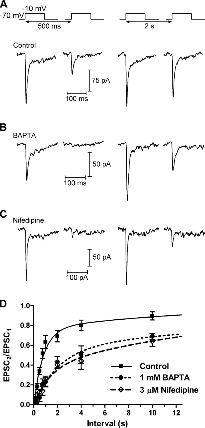
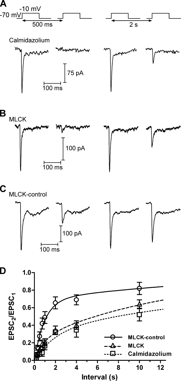
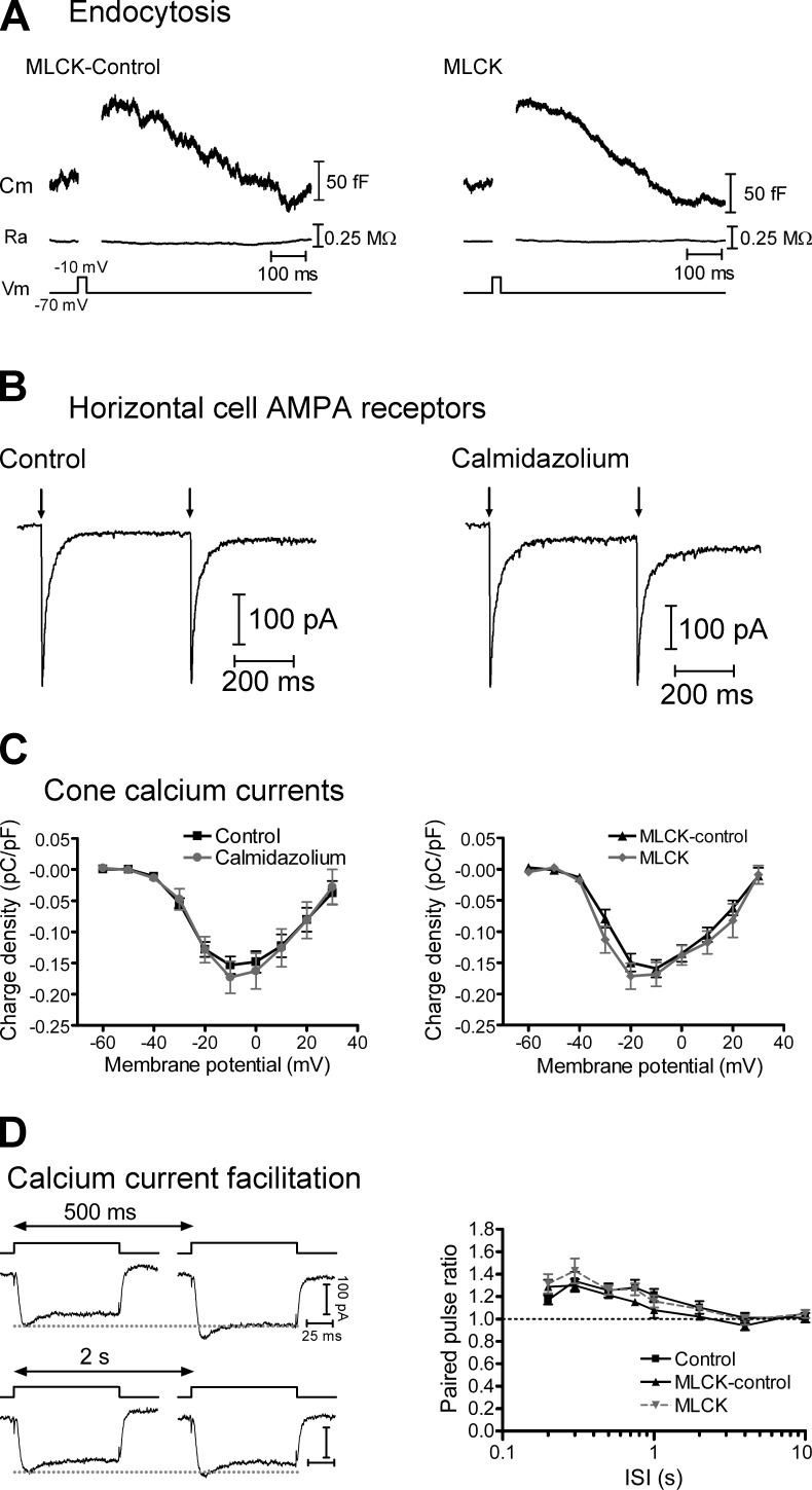
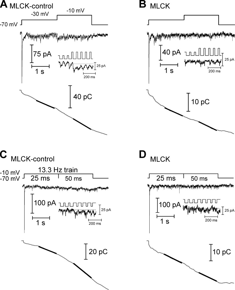
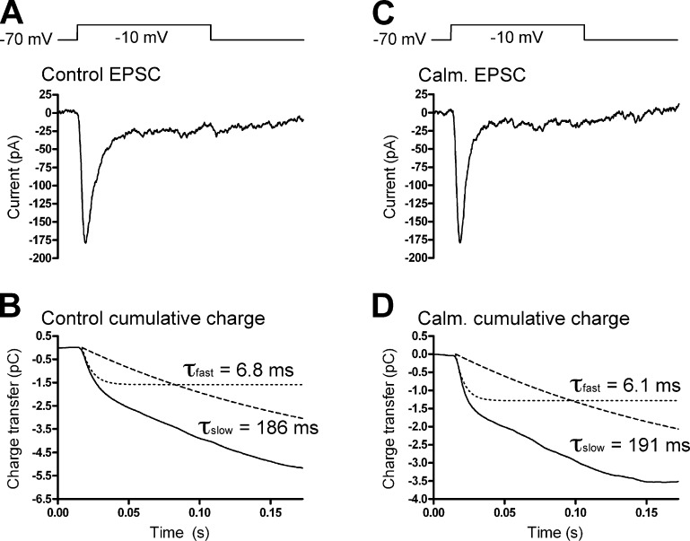
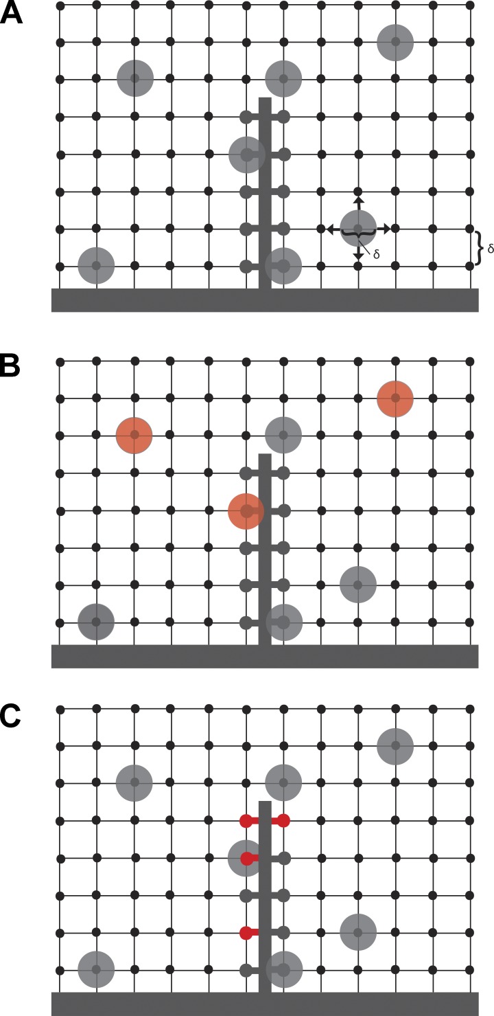
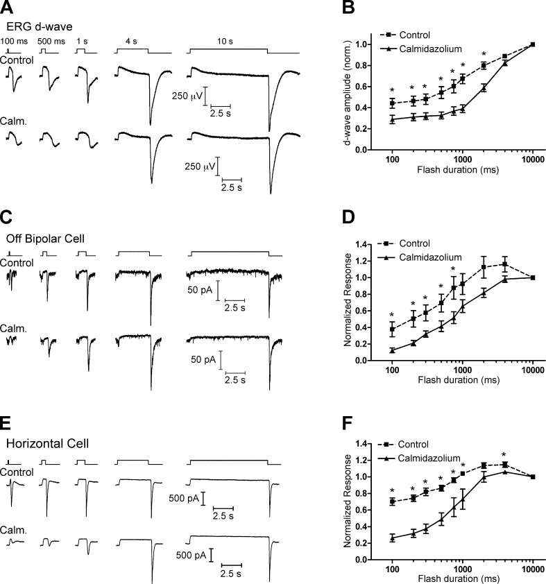
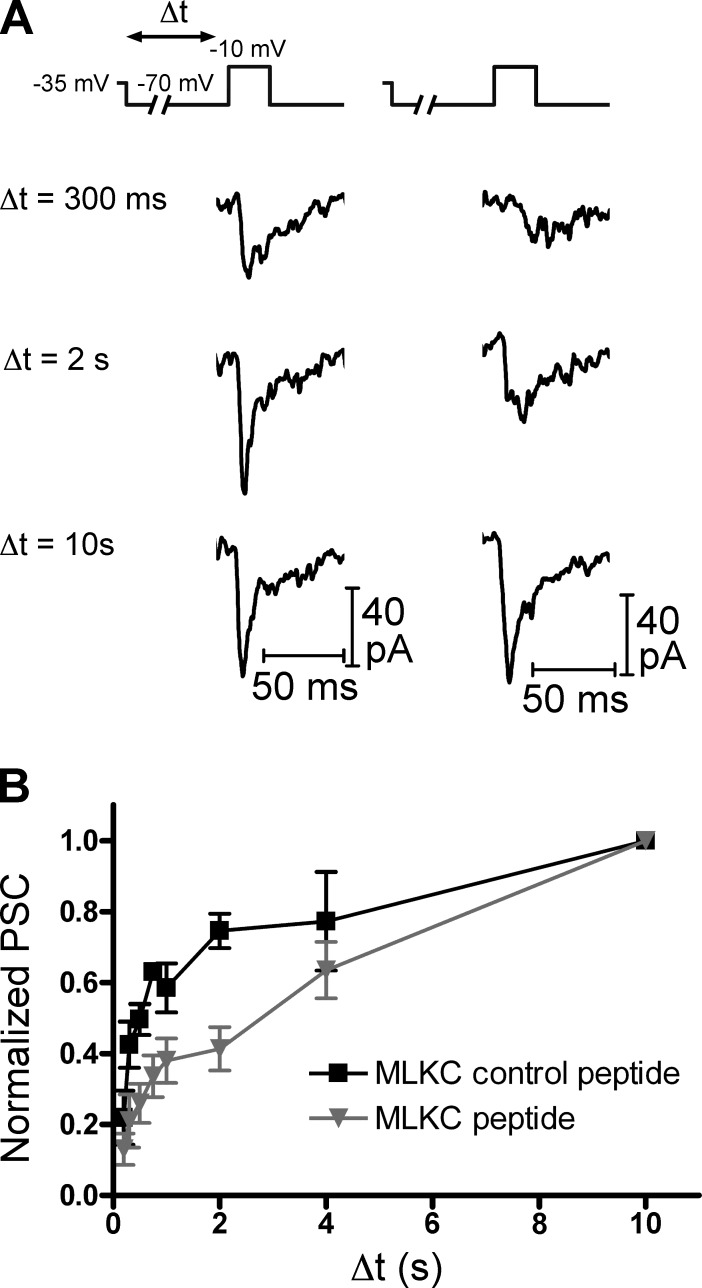
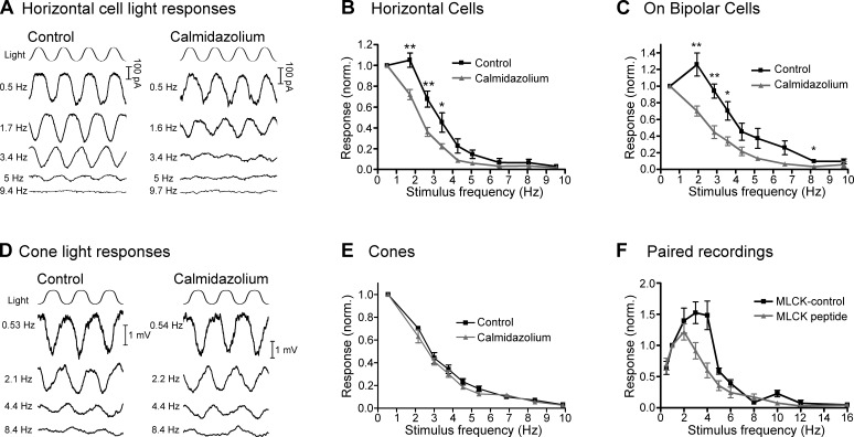
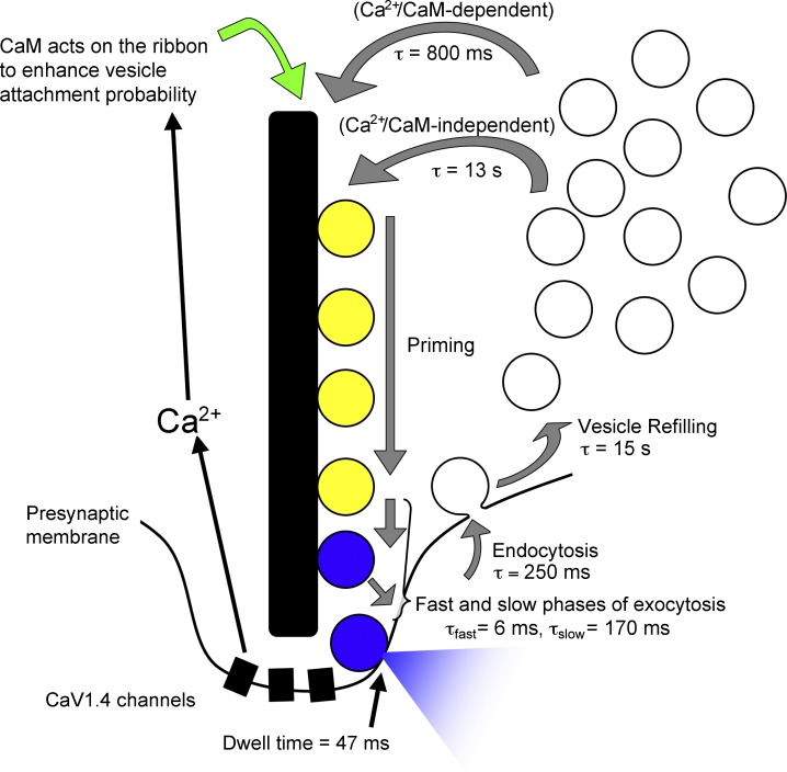
At the first synapse in the vertebrate visual pathway, light-evoked changes in photoreceptor membrane potential alter the rate of glutamate release onto second-order retinal neurons. This process depends on the synaptic ribbon, a specialized structure found at various sensory synapses, to provide a supply of primed vesicles for release. Calcium (Ca(2+)) accelerates the replenishment of vesicles at cone ribbon synapses, but the mechanisms underlying this acceleration and its functional implications for vision are unknown. We studied vesicle replenishment using paired whole-cell recordings of cones and postsynaptic neurons in tiger salamander retinas and found that it involves two kinetic mechanisms, the faster of which was diminished by calmodulin (CaM) inhibitors. We developed an analytical model that can be applied to both conventional and ribbon synapses and showed that vesicle resupply is limited by a simple time constant, τ = 1/(Dρδs), where D is the vesicle diffusion coefficient, δ is the vesicle diameter, ρ is the vesicle density, and s is the probability of vesicle attachment. The combination of electrophysiological measurements, modeling, and total internal reflection fluorescence microscopy of single synaptic vesicles suggested that CaM speeds replenishment by enhancing vesicle attachment to the ribbon. Using electroretinogram and whole-cell recordings of light responses, we found that enhanced replenishment improves the ability of cone synapses to signal darkness after brief flashes of light and enhances the amplitude of responses to higher-frequency stimuli. By accelerating the resupply of vesicles to the ribbon, CaM extends the temporal range of synaptic transmission, allowing cones to transmit higher-frequency visual information to downstream neurons. Thus, the ability of the visual system to encode time-varying stimuli is shaped by the dynamics of vesicle replenishment at photoreceptor synaptic ribbons.


Figures

Figure 1.
Figure 1.
Recovery from synaptic depression depends on Ca2+. (A) In paired recordings, a 100-ms depolarization of a cone to −10 mV from a holding potential of −70 mV evoked an EPSC in an HC. A second depolarization applied after a short interval (500 ms) evoked a smaller EPSC. The amplitude of the second EPSC largely recovered after a 2-s interval. (B) When the cone pipette solution contained 1 mM BAPTA, which restricts the spread of intracellular [Ca2+] away from Ca2+ channels, the amplitude of the second EPSC was reduced relative to control conditions. (C) Subsaturating inhibition of L-type Ca2+ channels with bath-applied nifedipine (3 µM), which reduced Ca2+ influx by ∼40%, also inhibited recovery from paired pulse depression, similar to BAPTA. (D) Group data showing the recovery from synaptic depression. As the interval between pulses increased, the PPR of the EPSCs (EPSC2/EPSC1) could be fit with two exponentials. Under control conditions, the fast time constant (τfast) was 816 ms (76%) and the slow time constant (τslow) was 12.9 s. In the presence of 1 mM BAPTA or 3 µM nifedipine, recovery from synaptic depression was slowed; with BAPTA, τfast = 2.1 s (56%) and τslow = 28.8s. In the presence of nifedipine, τfast = 1.4 s (37%) and τslow = 17.6 s. Mean ± SEM is shown.
Figure 2.
Figure 2.
Recovery from synaptic depression depends on CaM. (A) In a paired recording of a cone and an HC, there was minimal recovery from synaptic depression after an interstimulus interval of 500 ms when the retinal slices were treated with the membrane-permeant CaM inhibitor calmidazolium (20 µM). The EPSC recovered only partially after an interval of 2 s. (B) Likewise, when cones were dialyzed with a CaM-inhibiting MLCK (20 µM), recovery from synaptic depression was inhibited. (C) Recovery from synaptic depression resembled controls (Fig. 1) when the cone was dialyzed with a control version of the MLCK peptide (MLCK-control; 20 µM). (D) Group data. Recovery from synaptic depression was slowed when CaM was inhibited. In the presence of calmidazolium, τfast = 1.98 s (33%) and τslow = 25.9s. With MLCK, τfast = 917 ms (23%) and τslow = 13.8 s. With MLCK-control, τfast = 951 ms (68.1%) and τslow = 17.6 s. Mean ± SEM is shown.
Figure 3.
Figure 3.
CaM inhibitors do not affect rapid endocytosis, HC glutamate receptors, or cone Ca2+ currents (ICa). (A) Whole-cell capacitance recordings from cones that were dialyzed with either the MLCK-control (left) or MLCK (right) peptides (20 µM). There was a brief increase in whole-cell capacitance in response to a 25-ms depolarizing step to −10 mV from a holding potential of −70 mV, resulting from fusion of synaptic vesicles. The capacitance then decayed back toward baseline as vesicle membrane was retrieved via endocytosis. There was no change in access resistance (Ra). (B) Pairs of HC AMPA receptor currents were evoked by uncaging of MNI-glutamate (1 mM) with short (1 ms) UV flashes (500-ms interval) in control conditions (left) and, in a second HC, in the presence of 20 µM calmidazolium (right). Neither the amplitude nor the PPR was altered by calmidazolium. (C) Charge-voltage plots of leak-subtracted cone ICa recorded in response to 100-ms steps from −60 to 30 mV from a holding potential of −70 mV. The charge was normalized to whole-cell capacitance. Neither the amplitude nor the voltage dependence of ICa was affected by calmidazolium (20 µM, left) or the MLCK peptide (20 µM, right). (D) The PPR of Ca2+ charge (QCa2/QCa1) facilitated in response to pairs of depolarizing pulses (steps to −10 mV from −70 mV, 100-ms duration with intervals of 200 ms to 10 s). This facilitation was unaffected when cones were dialyzed with the MLCK or MLCK-control peptides. Mean ± SEM is shown.
Figure 4.
Figure 4.
Ca2+-dependent acceleration of replenishment measured using trains of depolarizing pulses. (A) In a paired recording of a cone and HC in which the cone was dialyzed with the MLCK-control peptide (20 µM), the cone was stimulated with a train of depolarizing pulses to −30 mV (25-ms duration, 13.3 Hz), evoking an EPSC in the HC (top). After 2 s, the step amplitude was increased to −10 mV to fully activate the Ca2+ current, accelerating replenishment. The inset shows the EPSC at the transition from steps to −30 mV to steps to −10 mV. The trace at the bottom shows the cumulative charge transfer obtained by integrating the EPSC waveform. The slope of a line (black line) fit to the final 1 s of the cumulative charge transfer of the EPSC in each experimental condition provides a measure of the rate of replenishment. (B) In a similar experiment, the cone was dialyzed with the MLCK peptide (20 µM). This inhibited the Ca2+-dependent acceleration of replenishment when the step amplitude was increased. (C) In a cone-HC paired recording in which the cone was dialyzed with the MLCK-control peptide, replenishment was accelerated when the depolarizing step (−70 to −10 mV) was lengthened from 25 to 50 ms. (D) This acceleration was inhibited when a cone was instead dialyzed with the MLCK peptide.
Figure 5.
Figure 5.
Cone release kinetics. (A) EPSC recorded under control conditions in an HC in response to a 100-ms depolarization of a presynaptic cone. (B) The cumulative charge transfer of the EPSC was fit with two exponential functions, with τfast = 6.8 ms and τslow = 186 ms. (C and D) Recording from another cone-HC pair in the presence of calmidazolium (Calm.; 20 µM). Similar to the control recording, the cumulative charge transfer (D) was fit with two exponential functions with τfast = 6.1 ms and τslow = 191 ms.
Figure 6.
Figure 6.
Model of ribbon replenishment. (A) The terminal was modeled as a three-dimensional rectangular lattice of vesicle sites separated by δ, the diameter of a single vesicle (δ = 45 nm). This space was populated with vesicles at a density of ρ = 2234 v/µm3 that move through the terminal with a diffusion coefficient D = 0.11 µm2/s. The terminal also contains a synaptic ribbon, which has 110 vesicle attachment sites. (B and C) Two simple variations of the model in which CaM acts to alter the vesicle attachment probability by acting on either vesicles themselves (B, red) or vesicle attachment sites on the ribbon (C, red).
Figure 7.
Figure 7.
CaM-dependent vesicle replenishment enhances responses at light offset. (A) Intraretinal ERG recordings were made in a superfused eyecup preparation in the presence of 10 µM L-AP4 to isolate the d-wave, which is the negative-going peak occurring at light offset in these intraretinal ERG recordings. (top) The amplitude of the d-wave increased with increasing flash duration. (bottom) When the eyecup was bathed with 20 µM calmidazolium (Calm.), the d-wave amplitude was reduced after short flashes. (B) Group data showing the reduction in d-wave amplitude when the eyecup was treated with calmidazolium. (C) A similar effect was seen in whole-cell recordings from Off BCs in a retinal slice. (top) The amplitude of a fast inward current at light offset increased with increasing flash duration. (bottom) When the retinal slices were bathed with 20 µM calmidazolium, the response was reduced after shorter-duration flashes. (D) Group data of the normalized charge of the response at light offset (integrated over 500 ms after the end of the flash), showing that the off response in Off BCs was reduced in the presence of calmidazolium. (E and F) Similar flash-duration experiments conducted with whole-cell recordings of HCs. Mean ± SEM is shown. *, P < 0.05.
Figure 8.
Figure 8.
Simulated light flash experiments. (A) A light flash was mimicked in paired recordings of cones and HCs. The cone was voltage-clamped at −35 mV to mimic the dark potential and hyperpolarized to −70 mV for a variable duration (Δt) to mimic a strong light flash before being depolarized to −10 mV (25 ms) to deplete the rapidly releasing vesicle pool. Cones were dialyzed with pipette solutions containing either the MLCK-control (20 µM, left) or MLCK (20 µM, right) peptide. (B) When cones were dialyzed with the MLCK-control peptide, the EPSC amplitude increased with increasing Δt. The amplitude of the EPSC was reduced when cones were instead dialyzed with the MLCK peptide. EPSC amplitude was normalized to the EPSC after the 10-s duration step. Mean ± SEM is shown.
Figure 9.
Figure 9.
Synaptic responses to sinusoidal stimuli are reduced by inhibition of CaM. (A) In control voltage-clamp recordings of an HC (left), response amplitude to a sinusoidal light stimulus decreased with increasing stimulus frequency. In recordings from a different HC (right), response amplitude in the presence of 20 µM calmidazolium declined at lower frequencies relative to control recordings. (B) HC group data normalized to the response amplitude at 0.5 Hz. In the presence of calmidazolium, responses fell off at lower frequencies than in controls. (C) A similar effect was seen in recordings from On BCs. (D) Perforated patch current-clamp recordings of responses from two separate cones to sinusoidal light stimuli in control conditions (left) and in the presence of the CaM inhibitor calmidazolium (20 µM; right). (E) Under control conditions, the cone light responses were exclusively low pass, peaking at the lowest frequency tested (0.5 Hz) and falling off at higher frequencies. In the presence of calmidazolium, the responses were no different than in control conditions. (F) In paired recordings of cones and HCs, cones were stimulated with a 0.5–16-Hz sinusoidal voltage-clamp command (20 mV peak-to-peak around a holding potential of −40 mV). Data were normalized to responses at 1 Hz. When cones were dialyzed with the MLCK-control peptide (20 µM; n = 8), HC responses were band-pass, peaking at 3–4 Hz and falling off at higher or lower frequencies. When cones were dialyzed with the MLCK peptide (gray; 20 µM), response amplitude fell off at lower frequencies than in controls. Mean ± SEM is shown. *, P < 0.05; **, P < 0.01.
Figure 10.
Figure 10.
Proposed vesicle cycle at the cone ribbon synapse. Vesicles can be resupplied to the synaptic ribbon through a fast mechanism (τ ∼800 ms) that is regulated by the actions of Ca2+/CaM on vesicle attachment sites on the ribbon, several hundred nanometers distant from Ca2+ entry through channels located at the ribbon base. Vesicles can also attach through a slower, Ca2+/CaM-independent process that has a time constant of ∼13 s. Vesicle priming appears to involve the synaptic ribbon (Snellman et al., 2011). Exocytosis occurs in two phases, with time constants of 6 ms and 170 ms that represent fast fusion of the IRP (blue) and movement of vesicles from the ribbon-associated reserve pool (yellow) to release sites near the ribbon base, respectively. Endocytosis is fast (τ = 250 ms; Van Hook and Thoreson, 2012) and returns vesicles to a cytoplasmic reservoir pool, where they are refilled with glutamate (τ ∼15 s; Hori and Takahashi, 2012).

References

    1. Abbott, L.F., and Regehr W.G.. 2004. Synaptic computation. Nature. 431:796–803. 10.1038/nature03010 - DOI - PubMed
    1. Armstrong-Gold, C.E., and Rieke F.. 2003. Bandpass filtering at the rod to second-order cell synapse in salamander (Ambystoma tigrinum) retina. J. Neurosci. 23:3796–3806 - PMC - PubMed
    1. Awatramani, G., Wang J., and Slaughter M.M.. 2001. Amacrine and ganglion cell contributions to the electroretinogram in amphibian retina. Vis. Neurosci. 18:147–156. 10.1017/S0952523801181149 - DOI - PubMed
    1. Babai, N., Bartoletti T.M., and Thoreson W.B.. 2010. Calcium regulates vesicle replenishment at the cone ribbon synapse. J. Neurosci. 30:15866–15877. 10.1523/JNEUROSCI.2891-10.2010 - DOI - PMC - PubMed
    1. Barrow, A.J., and Wu S.M.. 2009. Low-conductance HCN1 ion channels augment the frequency response of rod and cone photoreceptors. J. Neurosci. 29:5841–5853. 10.1523/JNEUROSCI.5746-08.2009 - DOI - PMC - PubMed

Publication types