

Orchestration of pulmonary T cell immunity during Mycobacterium tuberculosis infection: immunity interruptus
- PMID: 25311810
- PMCID: PMC4250436
- DOI: 10.1016/j.smim.2014.09.003
Orchestration of pulmonary T cell immunity during Mycobacterium tuberculosis infection: immunity interruptus
Abstract
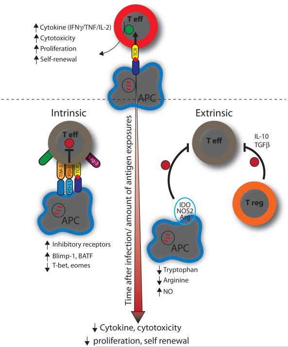
Despite the introduction almost a century ago of Mycobacterium bovis BCG (BCG), an attenuated form of M. bovis that is used as a vaccine against Mycobacterium tuberculosis, tuberculosis remains a global health threat and kills more than 1.5 million people each year. This is mostly because BCG fails to prevent pulmonary disease--the contagious form of tuberculosis. Although there have been significant advances in understanding how the immune system responds to infection, the qualities that define protective immunity against M. tuberculosis remain poorly characterized. The ability to predict who will maintain control over the infection and who will succumb to clinical disease would revolutionize our approach to surveillance, control, and treatment. Here we review the current understanding of pulmonary T cell responses following M. tuberculosis infection. While infection elicits a strong immune response that contains infection, M. tuberculosis evades eradication. Traditionally, its intracellular lifestyle and alteration of macrophage function are viewed as the dominant mechanisms of evasion. Now we appreciate that chronic inflammation leads to T cell dysfunction. While this may arise as the host balances the goals of bacterial sterilization and avoidance of tissue damage, it is becoming clear that T cell dysfunction impairs host resistance. Defining the mechanisms that lead to T cell dysfunction is crucial as memory T cell responses are likely to be subject to the same subject to the same pressures. Thus, success of T cell based vaccines is predicated on memory T cells avoiding exhaustion while at the same time not promoting overt tissue damage.
Keywords: Cytokine; Exhaustion; Memory; Priming; T cell; Tuberculosis.
Copyright © 2014 Elsevier Ltd. All rights reserved.
Figures
Similar articles
-
Pro- and anti-inflammatory cytokines in tuberculosis: a two-edged sword in TB pathogenesis.Semin Immunol. 2014 Dec;26(6):543-51. doi: 10.1016/j.smim.2014.09.011. Epub 2014 Nov 8. Semin Immunol. 2014. PMID: 25453229 Review.
-
Influence of Mycobacterium bovis BCG vaccination on cellular immune response of guinea pigs challenged with Mycobacterium tuberculosis.Clin Vaccine Immunol. 2008 Aug;15(8):1248-58. doi: 10.1128/CVI.00019-08. Epub 2008 May 28. Clin Vaccine Immunol. 2008. PMID: 18508930 Free PMC article.
-
B cells regulate neutrophilia during Mycobacterium tuberculosis infection and BCG vaccination by modulating the interleukin-17 response.PLoS Pathog. 2013;9(7):e1003472. doi: 10.1371/journal.ppat.1003472. Epub 2013 Jul 11. PLoS Pathog. 2013. PMID: 23853593 Free PMC article.
-
Critical and independent role for SOCS3 in either myeloid or T cells in resistance to Mycobacterium tuberculosis.PLoS Pathog. 2013;9(7):e1003442. doi: 10.1371/journal.ppat.1003442. Epub 2013 Jul 4. PLoS Pathog. 2013. PMID: 23853585 Free PMC article.
-
Understanding delayed T-cell priming, lung recruitment, and airway luminal T-cell responses in host defense against pulmonary tuberculosis.Clin Dev Immunol. 2012;2012:628293. doi: 10.1155/2012/628293. Epub 2012 Apr 1. Clin Dev Immunol. 2012. PMID: 22545059 Free PMC article. Review.
Cited by
-
Cytokine Receptors-Regulators of Antimycobacterial Immune Response.Int J Mol Sci. 2022 Jan 20;23(3):1112. doi: 10.3390/ijms23031112. Int J Mol Sci. 2022. PMID: 35163035 Free PMC article. Review.
-
Effect of bacillus Calmette-Guérin vaccination on CD4+Foxp3+ T cells during acquired immune response to Mycobacterium tuberculosis infection.J Leukoc Biol. 2016 Apr;99(4):605-17. doi: 10.1189/jlb.4A0614-308RR. Epub 2015 Nov 20. J Leukoc Biol. 2016. PMID: 26590147 Free PMC article.
-
The Frequency and Effect of Granulocytic Myeloid-Derived Suppressor Cells on Mycobacterial Survival in Patients With Tuberculosis: A Preliminary Report.Front Immunol. 2021 Jun 1;12:676679. doi: 10.3389/fimmu.2021.676679. eCollection 2021. Front Immunol. 2021. PMID: 34149712 Free PMC article.
-
Influence of the polymorphism of the DUSP14 gene on the expression of immune-related genes and development of pulmonary tuberculosis.Genes Immun. 2016 Jun;17(4):207-12. doi: 10.1038/gene.2016.11. Epub 2016 Mar 3. Genes Immun. 2016. PMID: 26938665
-
T Cells Primed by Live Mycobacteria Versus a Tuberculosis Subunit Vaccine Exhibit Distinct Functional Properties.EBioMedicine. 2018 Jan;27:27-39. doi: 10.1016/j.ebiom.2017.12.004. Epub 2017 Dec 7. EBioMedicine. 2018. PMID: 29249639 Free PMC article.
References
-
- Horsburgh CR, Jr., Rubin EJ. Latent Tuberculosis Infection in the United States. N Engl J Med. 2011;364:1441–8. - PubMed
-
- North RJ, Jung Y-J. Immunity to tuberculosis. Annu Rev Immunol. 2004;22:599–623. - PubMed
-
- Tian T, Woodworth J, Sköld M, Behar S. In vivo depletion of CD11c+ cells delays the CD4+ T cell response to Mycobacterium tuberculosis and exacerbates the outcome of infection. J Immunol. 2005;175:3268–72. - PubMed
-
- Hanekom WA, Mendillo M, Manca C, Haslett PAJ, Siddiqui MR, Barry C, et al. Mycobacterium tuberculosis inhibits maturation of human monocyte-derived dendritic cells in vitro. J Infect Dis. 2003;188:257–66. - PubMed
Publication types
MeSH terms
Substances
Grants and funding
- R01 HL080312/HL/NHLBI NIH HHS/United States
- AI100766/AI/NIAID NIH HHS/United States
- AI085669/AI/NIAID NIH HHS/United States
- AI106725/AI/NIAID NIH HHS/United States
- AI098637/AI/NIAID NIH HHS/United States
- R01 AI085669/AI/NIAID NIH HHS/United States
- R01 HL080330/HL/NHLBI NIH HHS/United States
- R21 AI100766/AI/NIAID NIH HHS/United States
- ImNIH/Intramural NIH HHS/United States
- R01 AI106725/AI/NIAID NIH HHS/United States
- R01 AI067731/AI/NIAID NIH HHS/United States
- R56 AI067731/AI/NIAID NIH HHS/United States
- R01 AI098637/AI/NIAID NIH HHS/United States
LinkOut - more resources
Full Text Sources
Other Literature Sources
