

Gallbladder lymphoma in a miniature dachshund
- PMID: 25311915
- PMCID: PMC4349549
- DOI: 10.1292/jvms.14-0300
Gallbladder lymphoma in a miniature dachshund
Abstract
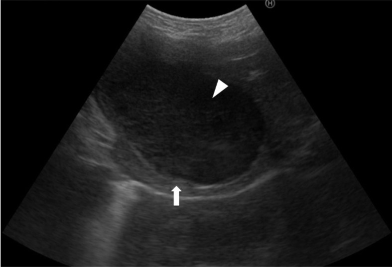
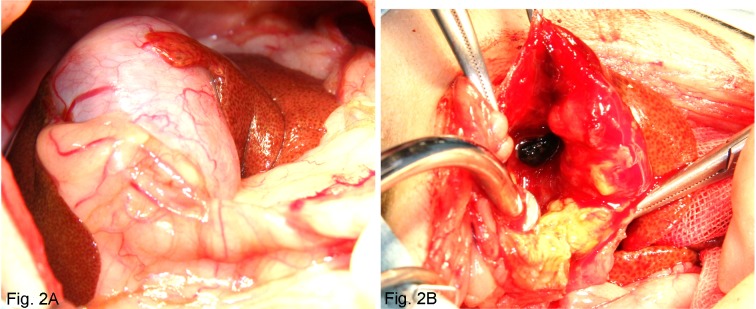
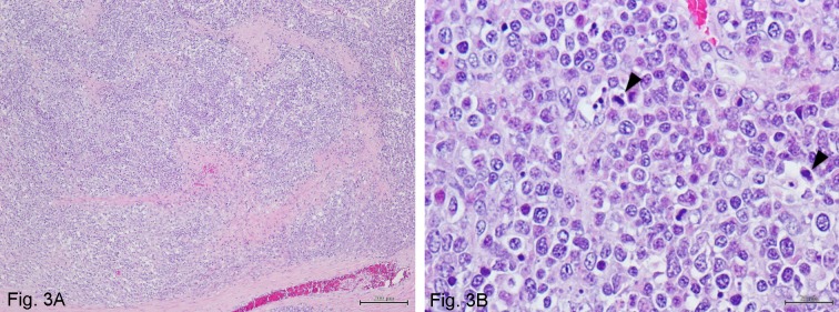
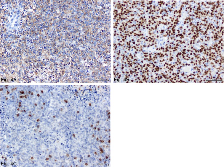
A 7-year-old, miniature dachshund was referred for examination and treatment of persistent anorexia, deep yellow-coloured urine and leucocytosis. The clinical sign of jaundice, results from a serum biochemistry profile and ultrasonographic images suggested a biliary tract obstruction. A cholecystectomy was performed to remove the obstruction. Histopathological assessment of the resected gallbladder and partial common bile duct indicated diffuse large B-cell lymphoma. Twelve days after the initial operation, a second procedure was performed due to bile leakage into the abdominal cavity. Chemotherapy was administered twice after the second operation but discontinued, because the dog showed adverse effects. The dog is still alive 24 months after the surgery. To the authors' knowledge, this is the first description of canine gallbladder lymphoma.
Figures
Similar articles
-
Clinicopathologic findings of splenic marginal zone lymphoma with gallbladder involvement that progressed to diffuse large B-cell lymphoma in a dog.Vet Clin Pathol. 2024 Jun;53(2):234-241. doi: 10.1111/vcp.13331. Epub 2024 May 28. Vet Clin Pathol. 2024. PMID: 38806404
-
Clinical outcomes of 30 cases (1997-2004) of canine gastrointestinal lymphoma.J Am Anim Hosp Assoc. 2007 Nov-Dec;43(6):313-21. doi: 10.5326/0430313. J Am Anim Hosp Assoc. 2007. PMID: 17975213
-
Rectal lymphoma in 11 dogs: a retrospective study.J Small Anim Pract. 2012 Oct;53(10):586-91. doi: 10.1111/j.1748-5827.2012.01258.x. Epub 2012 Aug 8. J Small Anim Pract. 2012. PMID: 22882127
-
Gastric Intravascular Lymphoma in a Dog: Case Report and Literature Review.J Am Anim Hosp Assoc. 2020 May/Jun;56(3):185. doi: 10.5326/JAAHA-MS-6994. Epub 2020 Mar 17. J Am Anim Hosp Assoc. 2020. PMID: 32182111 Review.
-
Treatment of lymphoma in dogs.J Am Vet Med Assoc. 1990 Mar 1;196(5):774-81. J Am Vet Med Assoc. 1990. PMID: 2407706 Review. No abstract available.
Cited by
-
Chronic Cholecystitis of Dogs: Clinicopathologic Features and Relationship with Liver.Animals (Basel). 2021 Nov 21;11(11):3324. doi: 10.3390/ani11113324. Animals (Basel). 2021. PMID: 34828055 Free PMC article.
References
-
- Bisig B., Copie-Bergman C., Baia M., Gaulard P., Delbecque K., Fassotte M. F., Boniver J., de Leval L.2009. Primary mucosa-associated lymphoid tissue lymphoma of the gallbladder: report of a case harboring API2/MALT1 gene fusion. Hum. Pathol. 40: 1504–1509. doi: 10.1016/j.humpath.2009.02.018 - DOI - PubMed
-
- Ferluga D., Luzar B., Gadzijev E. M.2003. Follicular lymphoma of the gallbladder and extrahepatic bile ducts. Virchows Arch. 442: 136–140. - PubMed
Publication types
MeSH terms
Substances
LinkOut - more resources
Full Text Sources
Other Literature Sources
Medical