. 2015 Feb;122(2):326-34.
doi: 10.1016/j.ophtha.2014.08.012. Epub 2014 Oct 12.

Clinical and molecular characteristics of childhood-onset Stargardt disease

Affiliations

Clinical and molecular characteristics of childhood-onset Stargardt disease

Kaoru Fujinami et al. Ophthalmology. 2015 Feb.

Abstract

Purpose: To describe the clinical and molecular characteristics of patients with childhood-onset Stargardt disease (STGD).

Design: Retrospective case series.

Participants: Forty-two patients who were diagnosed with STGD in childhood at a single institution between January 2001 and January 2012.

Methods: A detailed history and a comprehensive ophthalmic examination were undertaken, including color fundus photography, autofluorescence imaging, spectral-domain optical coherence tomography (SD-OCT), and pattern and full-field electroretinograms. The entire coding region and splice sites of ABCA4 were screened using a next-generation, sequencing-based strategy. The molecular genetic findings of childhood-onset STGD patients were compared with those of adult-onset patients.

Main outcome measures: Clinical, imaging, electrophysiologic, and molecular genetic findings.

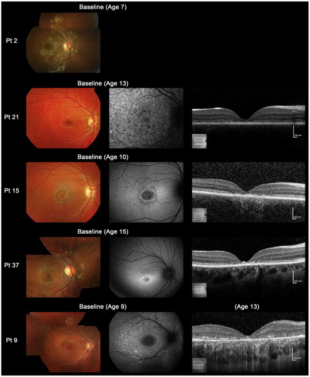
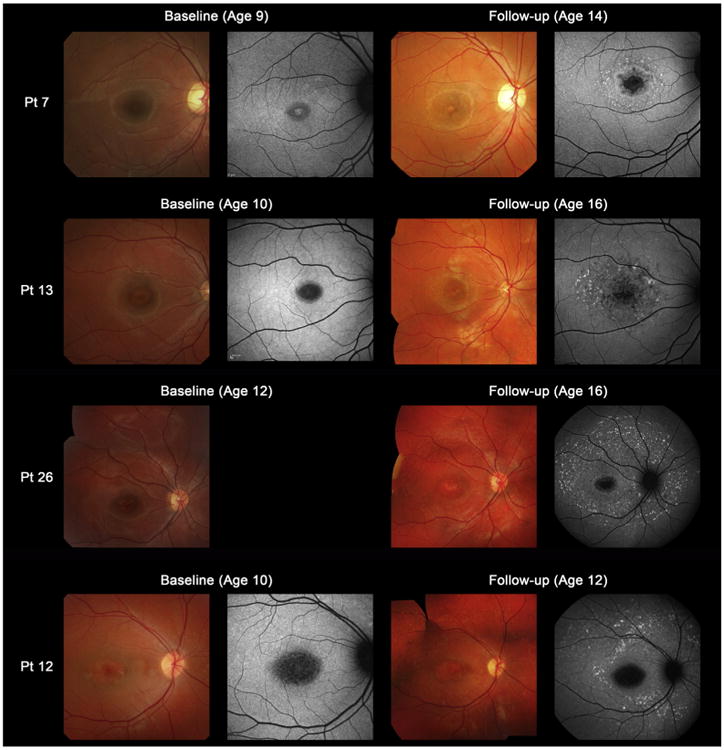
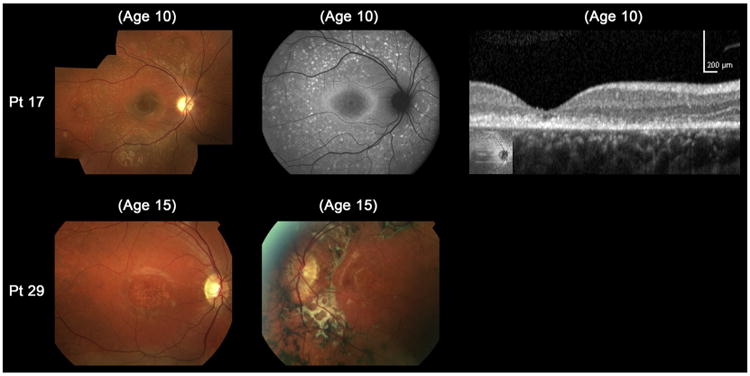
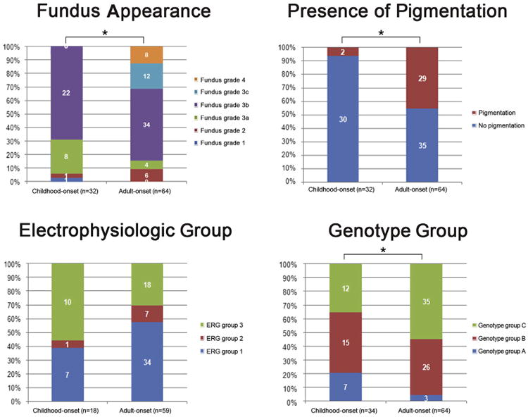
Results: The median ages of onset and the median age at baseline examination were 8.5 (range, 3-16) and 12.0 years (range, 7-16), respectively. The median baseline logarithm of the minimum angle of resolution visual acuity was 0.74. At baseline, 26 of 39 patients (67%) with available photographs had macular atrophy with macular/peripheral flecks; 11 (28%) had macular atrophy without flecks; 1 (2.5%) had numerous flecks without macular atrophy; and 1 (2.5%) had a normal fundus appearance. Flecks were not identified at baseline in 12 patients (31%). SD-OCT detected foveal outer retinal disruption in all 21 patients with available images. Electrophysiologic assessment demonstrated retinal dysfunction confined to the macula in 9 patients (36%), macular and generalized cone dysfunction in 1 subject (4%), and macular and generalized cone and rod dysfunction in 15 individuals (60%). At least 1 disease-causing ABCA4 variant was identified in 38 patients (90%), including 13 novel variants; ≥2 variants were identified in 34 patients (81%). Patients with childhood-onset STGD more frequently harbored 2 deleterious variants (18% vs 5%) compared with patients with adult-onset STGD.

Conclusions: Childhood-onset STGD is associated with severe visual loss, early morphologic changes, and often generalized retinal dysfunction, despite often having less severe fundus abnormalities on examination. One third of children do not have flecks at presentation. The relatively high proportion of deleterious ABCA4 variants supports the hypothesis that earlier onset disease is often owing to more severe variants in ABCA4 than those found in adult-onset disease.


Figures

Figure 1
Figure 1
Color fundus photographs, autofluorescence, and spectral-domain optical coherence tomographic images of 5 representative cases with childhood-onset Stargardt Disease (patients 2, 21, 15, 37, and 9). Color fundus photographs of patient 2 shows normal findings at age 7 (fundus grade: 1). Patient 21 has numerous flecks at the posterior pole without central atrophy (fundus grade: 2) and autofluorescence (AF) imaging demonstrates widespread multiple foci of high and low AF signal at the posterior pole with a heterogeneous background (AF type 2). Spectral-domain optical coherence tomography (SD-OCT) identifies marked outer retinal loss at the central macula. Patient 15 has central atrophy without flecks (fundus grade: 3a) and AF imaging demonstrates a localized low AF signal at the fovea with a high signal edge surrounded by a homogeneous background (AF type: 1). SD-OCT detects marked outer retinal loss at the central macula. Patient 37 has central atrophy with macular flecks (fundus grade: 3b) and a localized low AF signal at the fovea surrounded by a homogeneous background with perifoveal foci of high signal (AF type: 1). SD-OCT shows outer retinal loss at the central macula. Patient 9 has central atrophy with peripheral flecks extending anterior to the vascular arcades (fundus grade: 3b) and a localized low AF signal at the macula surrounded by a heterogeneous background and widespread foci of high AF signal extending anterior to the vascular arcades (AF type: 2). SD-OCT reveals outer retinal disruption at the macula. Pt = patient.
Figure 2
Figure 2
Color fundus photographs and autofluorescence (AF) images of 4 representative cases developing macular flecks during follow-up (patients 7, 13, 26, and 12). Color photograph of patient 7 at baseline shows subtle central atrophy without flecks (fundus grade 3a). At baseline, AF imaging demonstrates a localized low AF signal surrounded by an irregular high signal (AF type 1). Five years later, there is marked central atrophy with visible macular flecks (fundus grade 3b) and AF imaging demonstrates a localized low AF signal at the fovea with perifoveal foci of high signal (AF type 1). Patient 13 shows central atrophy with no visible flecks at baseline (fundus grade 3a), with AF imaging showing a localized low AF signal surrounded by subtle foci of high AF signal at the macula (AF type 1). Six years later, there are marked and increased macular flecks, also clearly seen on AF imaging (fundus grade 3b; AF type 1). Patient 26 has central atrophy with no visible flecks at baseline (fundus grade 3a), but marked flecks corresponding to foci of high signal on AF imaging are present 4 years later (fundus grade 3b; AF type 2). Patient 12 shows central atrophy with early subtle peripheral flecks at baseline (fundus grade 3b) and AF imaging demonstrates a localized low AF signal with subtle foci of high AF signal extending anterior to the vascular arcades (AF type 2). Two years later, there are marked and increased macular and peripheral flecks, which are also well-defined on AF imaging (fundus grade 3b; AF type 2). Pt = patient.
Figure 3
Figure 3
Color fundus photographs, autofluorescence (AF), and spectral-domain optical coherence tomographic images of 2 molecularly proven cases with “atypical” clinical features of childhood-onset Stargardt Disease (patients 17 and 29). Color photograph of patient 17 shows fine dots at the central macula surrounded by numerous peripheral flecks and AF imaging demonstrates well-defined dots associated with a high signal at the central macula surrounded by a ring of increased AF signal and numerous foci with high and low signal extending to the peripheral retina. Outer retinal loss at the macula is present on SD-OCT. Patient 29 has asymmetric fundus findings with central atrophy and peripheral flecks in the right eye and macular atrophy with flecks, subretinal fibrosis, and hyperpigmentation at the level of the retinal pigment epithelium in the left eye. Pt = patient.
Figure 4
Figure 4
Comparison of the distribution of fundus appearances, presence of pigmentation, electrophysiologic group, and genotype group between a cohort with childhood-onset Stargardt disease and a group with adult-onset Stargardt disease. There are significant differences in terms of fundus appearance classification, presence of pigmentation, and genotype group classification (*P < 0.05). A higher proportion of patients with childhood-onset Stargardt disease are in electrophysiologic group 3 compared with adult-onset Stargardt disease, but this difference does not attain significance. ERG = electroretinography.

Similar articles

Cited by

References

    1. Michaelides M, Hunt DM, Moore AT. The genetics of inherited macular dystrophies. J Med Genet. 2003;40:641–50. - PMC - PubMed
    1. Allikmets R, Singh N, Sun H, et al. A photoreceptor cell-specific ATP-binding transporter gene (ABCR) is mutated in recessive Stargardt macular dystrophy. Nat Genet. 1997;15:236–46. - PubMed
    1. Fishman GA. Fundus flavimaculatus. A clinical classification. Arch Ophthalmol. 1976;94:2061–7. - PubMed
    1. Michaelides M, Chen LL, Brantley MA, Jr, et al. ABCA4 mutations and discordant ABCA4 alleles in patients and siblings with bull's-eye maculopathy. Br J Ophthalmol. 2007;91:1650–5. - PMC - PubMed
    1. Fujinami K, Lois N, Davidson AE, et al. A longitudinal study of Stargardt disease: clinical and electrophysiologic assessment, progression, and genotype correlations. Am J Ophthalmol. 2013;155:1075–88. - PubMed

Publication types

MeSH terms

Substances