Case Reports
. 2014 Oct 14:14:763.
doi: 10.1186/1471-2407-14-763.

Primary ectopic atypical meningioma in the renal hilum: a case report

Affiliations
Case Reports

Primary ectopic atypical meningioma in the renal hilum: a case report

Ye-qing Mao et al. BMC Cancer. .

Abstract

Background: Primary ectopic atypical meningioma involving the renal hilum is rare. This is, to our knowledge, only the second case report of a primary retroperitoneal meningioma and the first case of an atypical subtype in this location.

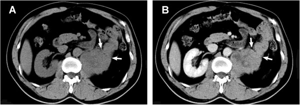
Case presentation: A 53-year-old Han Chinese man presented with a 2-year history of left-side flank pain. An oval-shaped retroperitoneal mass was found in the left renal hilum on computed tomography, which was resected en bloc along with the kidney via laparotomy. According to the World Health Organization criteria, the tumor was histopathologically classified as a meningioma (Grade II, atypical). Five years later, the tumor recurred at the primary site with a similar histopathology. The patient received palliative resection, followed by radiotherapy (4500 cGy in 25 fractions). No relapse was found at 6-month follow-up.

Conclusion: We describe the clinical, radiographic and histopathological features of an unusual case of aggressive ectopic meningioma in the renal hilum. The patient presented with a massive retroperitoneal tumor without primary cerebral or secondary metastatic lesions; the preoperative diagnosis was naturally confined to the common retroperitoneal malignancies. This case is of interest to oncologists, because of both its rare location and aggressiveness; it not only enriched the spectrum of primary ectopic meningioma, but also reminded us of potential recurrence of an atypical meningioma. This case raises the issue of the etiology of such a rare tumor that needs further investigation, and more importantly demands long-term follow-up result.


Figures

Figure 1
Figure 1
CT scanning and intravenous urography show the mass in the left renal hilum. The tumor displaced tissue near the renal hilum, and invaded the parenchyma and renal vessels. A: The plain scanning of the tumor. B: The contrast-enhanced phase of imaging. C: The collecting system of the affected kidney was compressed and distorted.
Figure 2
Figure 2
Recurrent mass in the renal fossa. A: CT scanning demonstrates that the tumor attached closely to the psoas muscle and invaded the jejunum (arrow). B: delayed, mild enhancement was prominent in the venous phase.
Figure 3
Figure 3
Images of small bowl obstruction. Gastrointestinal imaging showed a suspected obstruction at beginning of the jejunum.
Figure 4
Figure 4
Photomicrographs of histopathological examination. A: Necrotic foci appeared as an area filled with eosinophilic amorphous material (pentagram) [hematoxylin–eosin (HE)-stained, original magnification × 4]. B: Epithelioid spindle-shaped cells form in a storiform and sheet-like growth pattern (HE-stained, original magnification × 20). C: Fine structure of the tumor cells, and especially a typical mitotic figure (yellow arrow) are clearly demonstrated (HE-stained, original magnification × 40). D1: Immunohistochemical staining of EMA was positive (original magnification × 20). D2: Immunohistochemical staining of vimentin was positive (original magnification × 20). E: The Ki67 was estimated at 15%. F: Infiltration of the tumor (arrow) into longitudinal intestinal muscle layer (arrowhead; HE-stained, original magnification × 4).

References

    1. Claus EB, Morrison AL. Epidemiology of Meningiomas. In: DeMonte F, McDermott MW, Al-Mefty O, editors. Al-Mefty’s Meningioma. 2. New York: Thieme Medical; 2011. pp. 35–39.
    1. Huszar M, Fanburg JC, Dickersin GR, Kirshner JJ, Rosenberg AE. Retroperitoneal malignant meningioma: A light microscopic, immunohistochemical, and ultrastructural study. Am J Surg Pathol. 1996;20(4):492–499. doi: 10.1097/00000478-199604000-00014. - DOI - PubMed
    1. Ishigaki D, Arai H, Sasoh M, Ogasawara K, Uesugi N, Sugai T, Nakamura S, Ogawa A. Meningioma in the posterior fossa without dural attachment. Neurol Med Chir. 2007;47(8):364–366. doi: 10.2176/nmc.47.364. - DOI - PubMed
    1. Llauger J, Aixut S, Canete N, Palmer J, Sola M, Bague S. Meningioma of the scapula. Skeletal Radiol. 2008;37(2):169–171. doi: 10.1007/s00256-007-0366-7. - DOI - PubMed
    1. Lockett L, Chiang V, Scully N. Primary pulmonary meningioma: report of a case and review of the literature. Am J Surg Pathol. 1997;21(4):453–460. doi: 10.1097/00000478-199704000-00012. - DOI - PubMed
Pre-publication history
    1. The pre-publication history for this paper can be accessed here:http://www.biomedcentral.com/1471-2407/14/763/prepub

Publication types

LinkOut - more resources