. 2014 Nov 6;15(5):605-18.
doi: 10.1016/j.stem.2014.09.008. Epub 2014 Oct 9.

Bipotential adult liver progenitors are derived from chronically injured mature hepatocytes

Affiliations

Bipotential adult liver progenitors are derived from chronically injured mature hepatocytes

Branden D Tarlow et al. Cell Stem Cell. .

Abstract

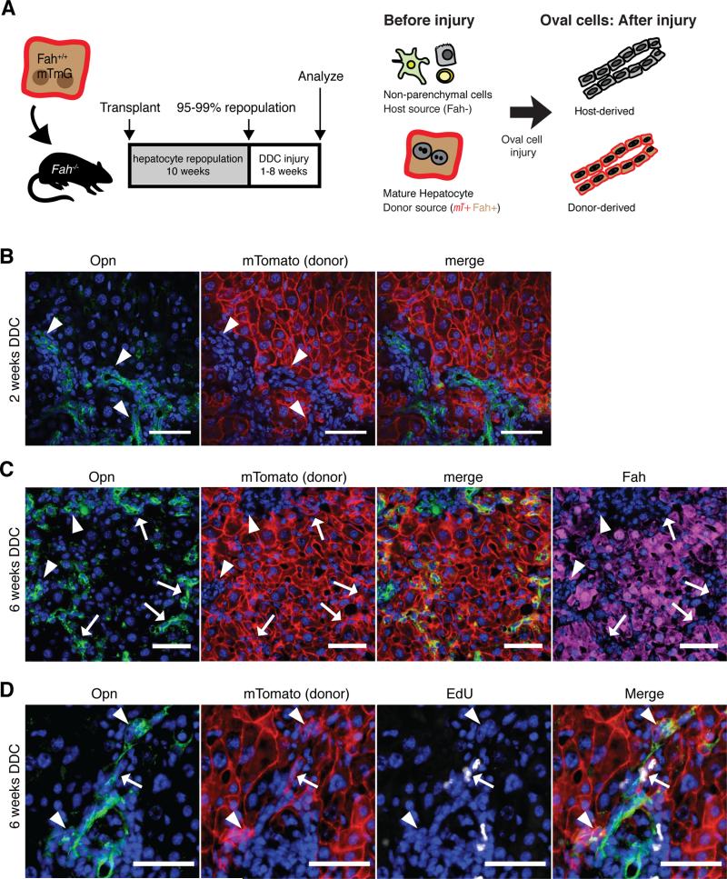
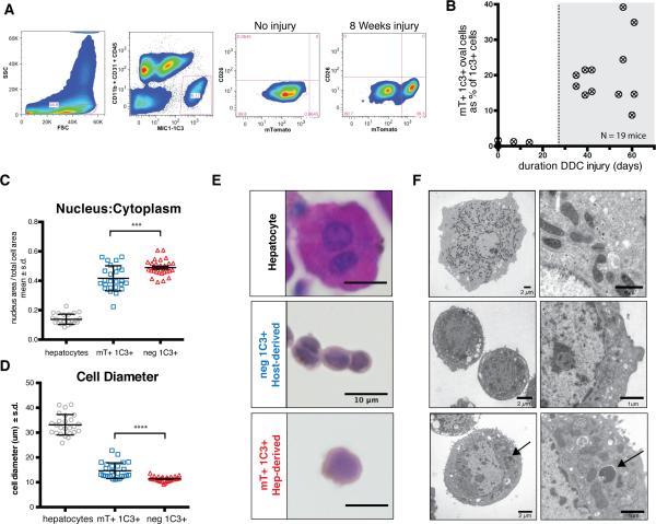
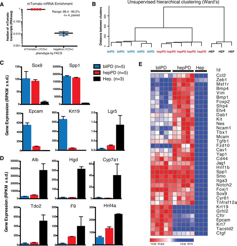
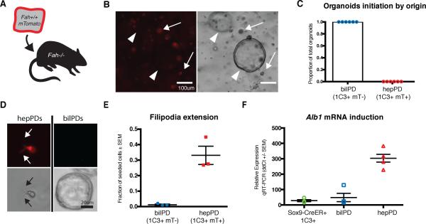
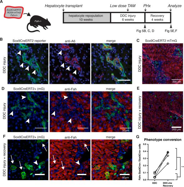
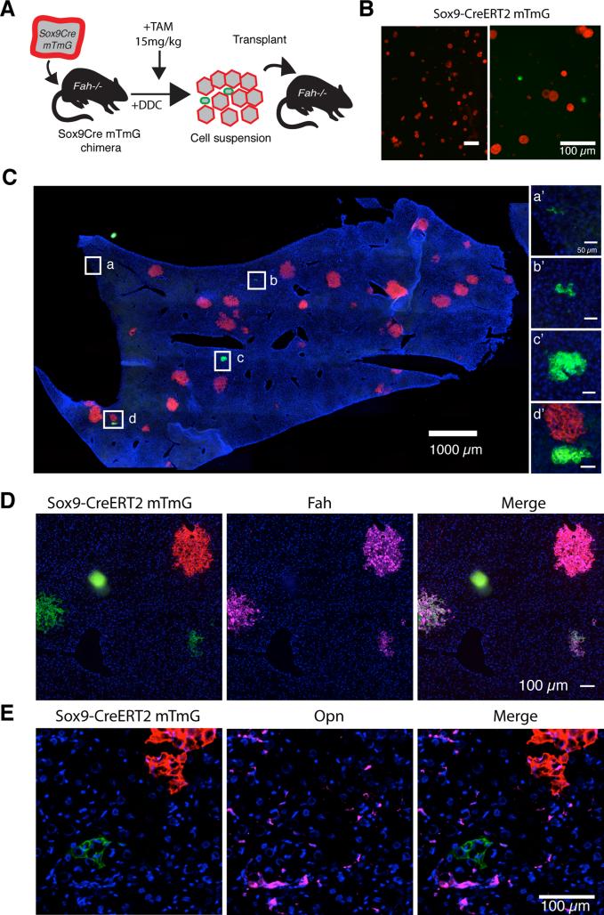
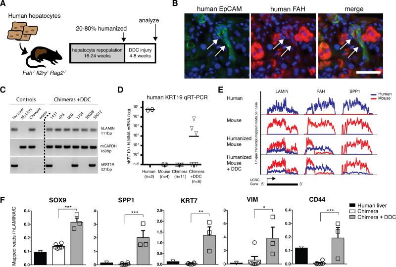
Adult liver progenitor cells are biliary-like epithelial cells that emerge only under injury conditions in the periportal region of the liver. They exhibit phenotypes of both hepatocytes and bile ducts. However, their origin and their significance to injury repair remain unclear. Here, we used a chimeric lineage tracing system to demonstrate that hepatocytes contribute to the progenitor pool. RNA-sequencing, ultrastructural analysis, and in vitro progenitor assays revealed that hepatocyte-derived progenitors were distinct from their biliary-derived counterparts. In vivo lineage tracing and serial transplantation assays showed that hepatocyte-derived proliferative ducts retained a memory of their origin and differentiated back into hepatocytes upon cessation of injury. Similarly, human hepatocytes in chimeric mice also gave rise to biliary progenitors in vivo. We conclude that human and mouse hepatocytes can undergo reversible ductal metaplasia in response to injury, expand as ducts, and subsequently contribute to restoration of the hepatocyte mass.


Figures

Figure 1
Figure 1. Hepatocyte-derived oval cells appear after extended injury
A) Purified hepatocytes fluorescently marked hepatocytes were transplanted into the spleen of Fah−/− animals. After 10 weeks repopulation, DDC injury was given for 1 to 8 weeks. Since only hepatocytes were marked at baseline, any fluorescent marked ductal cells observed after injury were inferred to be hepatocytes-derived. B) OPN+ ductal proliferation did not colocalize with hepatocyte marker mTomato after 2 weeks injury (arrowhead, bar = 50μm). C) After 6 weeks of injury, a subset of OPN+ ductal cells co- localized with hepatocyte derived mTomato marked cells (arrow), however, the majority of ductal proliferation was still host-derived (arrowhead). Induction of OPN correlated with the loss of FAH(arrows), bar = 50μm. D) Hepatocyte-derived progenitors (mTomato+ OPN+) incorporated EdU 6 hours after a pulse after 6 weeks of injury.
Figure 2
Figure 2. Hepatocyte-derived liver progenitors cells are isolated with MIC1-1C3 antibody
A) Dissociated livers were FACS sorted with gates applied for FSC/SSC (to include ductal cells, as shown), pulse width (not shown), PI (not shown), and MIC1-1C3+ CD11b CD31 CD45. MIC1-1C3+ cell were separated based on mTomato fluorescence (mature hepatocyte origin). Without injury mTomato+ cells were a trace component of MIC1-1C3+ population but increased with injury. B) The percentage of ductal cells derived from mTomato-marked hepatocytes is plotted against the number of days of DDC injury. Hepatocyte-derived MIC1-1C3+ ductal progenitors emerged after approximately 4 weeks injury. C) FACS isolated populations were fixed and nucleus to cytoplasmic ratios and D) cell diameter and were examined for each population (pairwise t-test, p<0.001 ***, p<0.0001****). E) Representative H&E staining (bars = 10μm) and F) transmission electron microscopy from directly isolated cells from each population (bar size indicated). The arrow indicates a membrane bound structure in a lysosome adjacent to mitochondria.
Figure 3
Figure 3. Hepatocyte-derived oval cells are transcriptional distinct from bile ducts
A) FACS separation of MIC1-1C3+ cells based on ROSA-mTomato resulted in 98.4-99.2% enrichment in mTomato+ cells relative to mTomato cells (paired analysis, n=4 animals). B) Unsupervised hierarchical clustering (Ward's method) of hepPDs (n=5) bilPDs (n=5), and hepatocytes (n=3). C) Gene expression levels (RPKM) for progenitor associated genes and D) Hepatocyte-associated genes (mean ± s.d.). E) Cluster analysis shows hepPDs express biliary progenitor associated genes and a distinctive mesenchymal signature.
Figure 4
Figure 4. hepPDs are functionally distinct in vitro
A) Fah−/− mice were repopulated with Fah+/+ Rosa-mTomato+ hepatocytes and injured with DDC. B) Organoids derived from crude non-parenchymal preps from DDC injured chimeric liver were seeded in matrigel for organoid formation. Hepatocyte-derived mTomato+ cells (arrows) did not initiate organoids. All organoids were mTomato (arrowhead). C) MIC1-1C3+ mTomato+ and mTomato- cells were seeded into matrigel for organoid formation. All organoids were host-derived (50-200 counted/animal, n=6 animals). D) hepPDs formed fillipodia (arrows) in matrigel while mTomato bilPDs formed spherical organoids. E) Fillipodia formation was quantified as a fraction of seeded cells. F) hepPDs cultured in hepatic differentiation medium induced albumin mRNA with 10.8-fold greater efficiency than bilPDs or MIC1-1C3+ cells from uninjured Sox9-CreERT2 reporter mice (unpaired t-test, p<0.001***).
Figure 5
Figure 5. hepPDs revert back to hepatocyctes in vivo
A) Fah+/+ ROSA-mTmG Sox9-CreERT2 hepatocytes were transplanted into Fah−/− recipients to generate chimeras. DDC injury was given to repopulated chimeras for 4 weeks, a low dose pulse of tamoxifen was given (15mg/kg), and injury was continued for 2 additional weeks. B) Tissue harvested by 1/3 partial hepatectomy showed most Sox9-CreERT2 marked (mGFP+) cells co-localized with A6 antigen (arrowhead). C) Low-power view shows Sox9-marked ductal cells in periportal zone. D) Sox9-CreERT2- marked cells have biliary morphology that do not co-localize with hepatocyte marker FAH. E) Following a 4-week recovery period, mGFP+ hepatocytes localized in the portal area. F) Upon healing Sox9-CreERT2 marked cells assumed hepatocyte morphology co-localized with FAH (arrow). G) Within animal comparison indicated that recovery from DDC injury was associated with a 5-fold increase in marked hepatocytes (6.6% versus 33.5%, p < 0.01**, paired t-test, n=4).
Figure 6
Figure 6. hepPDs differentiate back into hepatocytes upon serial transplantation
A) mGFP+ hepPD and mTomato+ hepatocytes were dissociated as single cells from DDC-injured chimeric mice for intrasplenic transplantation into Fah-/- mice. B) Sox9- CreERT2+ mGFP+ cells were smaller than hepatocytes and represented 6.3% of mTmG cells scored in 5 random fields before transplantation. C) After 5-weeks NTBC cycling, we assessed the rate at which mGFP+ hepPDs contributed to repopulation. mGFP+ clones were smaller than mTomato+ clones D) Hepatocyte-nodules expressed hepatocyte marker FAH but E) not biliary/progenitor marker OPN.
Figure 7
Figure 7. Human hepatocytes are directly converted into biliary-like cells in vivo
A) Human hepatocyte were transplanted into FRG mice. After 16-24 weeks repopulation, animals were fed 0.1% DDC for 4-8 weeks. B) After injury, human EpCAM+ FAHlow cells with ductal morphology emerged. C) KRT19 qRT-PCR assay on whole liver specifically amplified human KRT19. 3 of 6 DDC-treated chimeric mice had robust KRT19 induction. D) KRT19 levels relative to human LMNA were only 9-to-285 fold lower (n=3) than human liver reference samples (n=2). E) RNA-sequencing of whole chimeric livers effectively separated human (blue) from mouse (red) transcripts, graphed as unique transcript-mapped reads per position across each UCSC gene model (3 examples shown). The FAH transcript in chimeric livers shows expected truncation of mouse FahΔexon5 and non-sense mediated decay but full length human FAH. F) Human mRNA levels of ductal progenitor genes were quantified relative to housekeeping gene LMNA in normal human liver (n=1), chimeric livers (n=6), and DDC injured chimeric livers (n=3)(mean ± SEM, *p <0.05, **p < 0.01, *** p<0.001).

Comment in

References

    1. Alison MR, Golding M, Sarraf CE, Edwards RJ, Lalani EN. Liver damage in the rat induces hepatocyte stem cells from biliary epithelial cells. Ygast. 1996;110:1182–1190. - PubMed
    1. Azuma H, Paulk N, Ranade A, Dorrell C, Al-Dhalimy M, Ellis E, Strom S, Kay MA, Finegold M, Grompe M. Robust expansion of human hepatocytes in Fah−/−/Rag2−/−/Il2rg−/− mice. Nat Biotechnol. 2007;25:903–910. - PMC - PubMed
    1. Li Bin, Zheng Y-W, Sano Y, Taniguchi H. Evidence for Mesenchymal–Epithelial Transition Associated with Mouse Hepatic Stem Cell Differentiation. PLoS ONE. 2011;6:e17092. - PMC - PubMed
    1. Chen Y, Wong PP, Sjeklocha L, Steer CJ, Sahin MB. Mature hepatocytes exhibit unexpected plasticity by direct dedifferentiation into liver progenitor cells in culture. Hepatology. 2012;55:563–574. - PMC - PubMed
    1. Demetris AJ, Seaberg EC, Wennerberg A, Ionellie J, Michalopoulos GK. Ductular reaction after submassive necrosis in humans. Special emphasis on analysis of ductular hepatocytes. Am J Pathol. 1996;149:439–448. - PMC - PubMed

Publication types

Associated data