. 2015 Jan;35(1):41-51.
doi: 10.1128/MCB.01107-14. Epub 2014 Oct 13.

Immune cell inhibition by SLAMF7 is mediated by a mechanism requiring src kinases, CD45, and SHIP-1 that is defective in multiple myeloma cells

Affiliations

Immune cell inhibition by SLAMF7 is mediated by a mechanism requiring src kinases, CD45, and SHIP-1 that is defective in multiple myeloma cells

Huaijian Guo et al. Mol Cell Biol. 2015 Jan.

Abstract

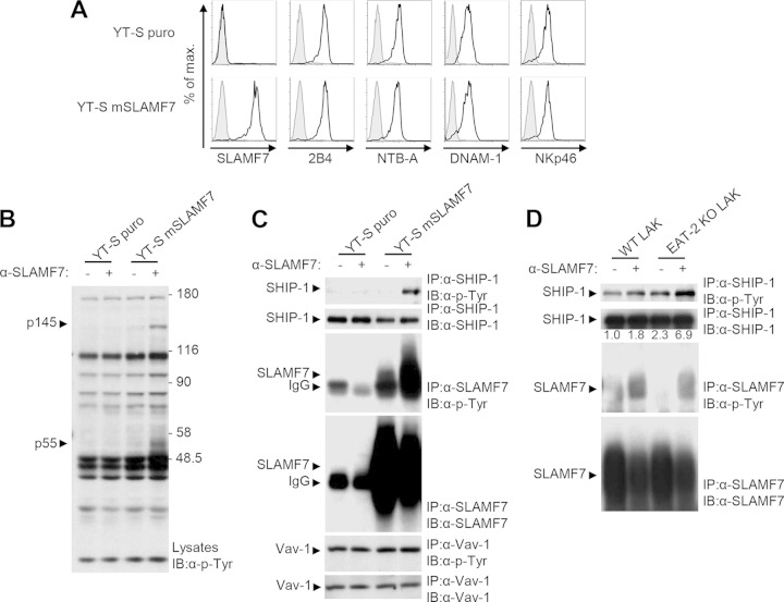
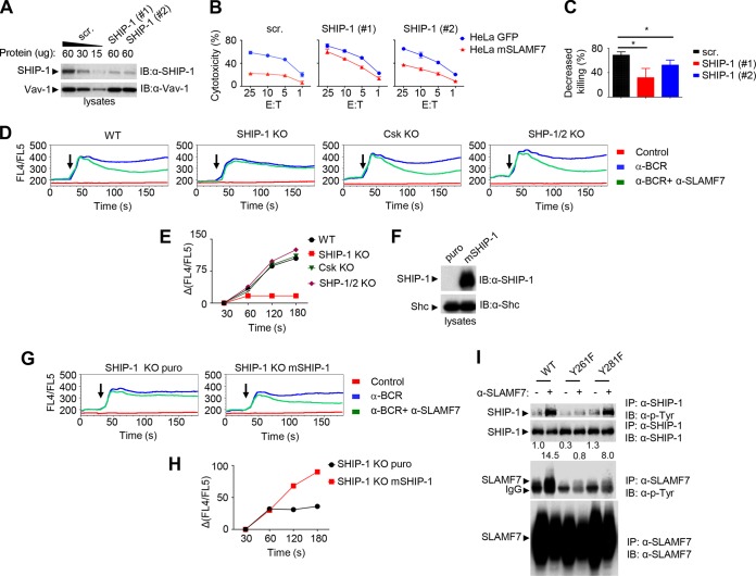
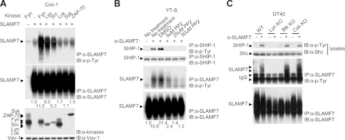
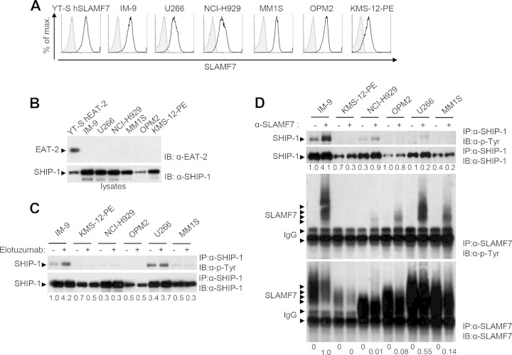
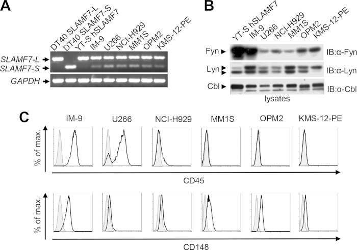
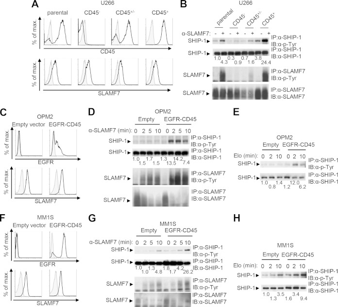
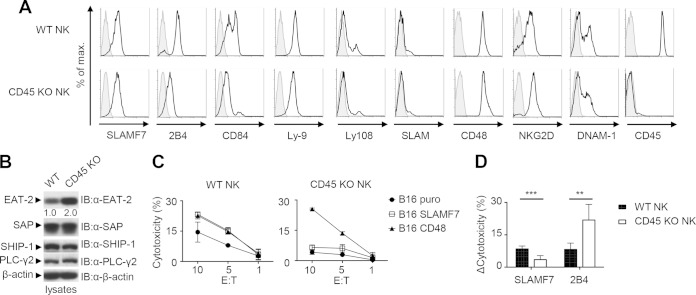
Signaling lymphocytic activation molecule F7 (SLAMF7) is a receptor present on immune cells, including natural killer (NK) cells. It is also expressed on multiple myeloma (MM) cells. This led to development of an anti-SLAMF7 antibody, elotuzumab, showing efficacy against MM. SLAMF7 mediates activating or inhibitory effects in NK cells, depending on whether cells express or do not express the adaptor EAT-2. Since MM cells lack EAT-2, we elucidated the inhibitory effectors of SLAMF7 in EAT-2-negative NK cells and tested whether these effectors were triggered in MM cells. SLAMF7-mediated inhibition in NK cells lacking EAT-2 was mediated by SH2 domain-containing inositol phosphatase 1 (SHIP-1), which was recruited via tyrosine 261 of SLAMF7. Coupling of SLAMF7 to SHIP-1 required Src kinases, which phosphorylated SLAMF7. Although MM cells lack EAT-2, elotuzumab did not induce inhibitory signals in these cells. This was at least partly due to a lack of CD45, a phosphatase required for Src kinase activation. A defect in SLAMF7 function was also observed in CD45-deficient NK cells. Hence, SLAMF7-triggered inhibition is mediated by a mechanism involving Src kinases, CD45, and SHIP-1 that is defective in MM cells. This defect might explain why elotuzumab eliminates MM cells by an indirect mechanism involving the activation of NK cells.


Figures

FIG 1
FIG 1
SHIP-1 is tyrosine phosphorylated in response to the engagement of SLAMF7. (A to C) YT-S cells were transfected with a vector encoding the puromycin resistance marker (puro) alone or in combination with mouse SLAMF7 (mSLAMF7). Polyclonal populations were used for analyses. (A) Expression of SLAMF7, 2B4, NTB-A, DNAM-1, and NKp46 (black lines with no shading) was analyzed by flow cytometry. Isotype controls are shown as gray lines with shading. Results are representative of 3 experiments. (B) Cells were stimulated or not stimulated for 3 min with the anti-SLAMF7 MAb 4G2 and rabbit anti-rat IgG. After lysis, overall protein tyrosine phosphorylation was determined by immunoblotting (IB) of total cell lysates with antiphosphotyrosine (α-p-Tyr) antibodies. The positions of prestained molecular mass markers (in kilodaltons) are shown on the right. (C) Tyrosine phosphorylation of SHIP-1, SLAMF7, and Vav-1 was determined by immunoprecipitating (IP) the indicated proteins with specific antibodies and subsequent immunoblotting with antiphosphotyrosine antibodies. The abundance of SHIP-1, SLAMF7, and Vav-1 in the immunoprecipitates was verified by probing parallel immunoprecipitates or reprobing the immunoblot membrane with substrate-specific antibodies. It is noteworthy that the amount of SLAMF7 recovered from cells stimulated with anti-SLAMF7 was frequently lower than that obtained from unstimulated cells. This was due to the fact that a proportion of SLAMF7 becomes detergent insoluble upon SLAMF7 stimulation with antibodies. The position of the heavy chain of IgG, which is recognized by the immunoblotting antibodies, is shown by an arrowhead on the left. Results are representative of 3 experiments. (D) NK cells from wild-type (WT) or EAT-2-deficient (knockout; KO) mice were stimulated for 3 min with anti-SLAMF7, and tyrosine phosphorylation of SHIP-1 and SLAMF7 was analyzed as detailed for panels A to C. A quantitation of the relative tyrosine phosphorylation of SHIP-1 is shown. Results are representative of 2 experiments.
FIG 2
FIG 2
SHIP-1 is required for SLAMF7-mediated inhibition in NK cells and DT40 B cells. (A to C) Expression of SHIP-1 in YT-S cells expressing mSLAMF7 was downregulated using two different SHIP-1-specific small interfering RNAs (siRNAs), #1 and #2. (A) Expression of SHIP-1 was determined by immunoblotting of total cell lysates with anti-SHIP-1 antibodies. Vav-1 was used as loading control. For cells transfected with scrambled siRNAs (scr.), various amounts of lysates were loaded to confirm the linearity of the immunoblot assays. (B) Cytotoxicity to HeLa target cells expressing green fluorescent protein (GFP) alone or in combination with mSLAMF7 was determined using a 51Cr release assay. The effector-to-target ratios (E:T) are shown at the bottom. Average values of duplicates are shown, while error bars depict standard deviations. Results are representative of 3 experiments. (C) The percentage of decreased killing was calculated as [(cytotoxicity to GFP+ targets − cytotoxicity to SLAMF7+ targets)/cytotoxicity to GFP+ targets] × 100 at the 25:1 E/T ratio. Average values from 3 independent experiments with standard deviations are represented. *, P < 0.05. (D and E) Wild-type (WT) DT40 cells or variants lacking the indicated inhibitory molecules were transduced with a cDNA encoding mouse SLAMF7. Cells were loaded with the Ca2+ indicator dye Indo-1 and then stimulated with anti-B cell receptor (α-BCR) antibodies alone or in combination with anti-SLAMF7. (D) Changes in intracellular Ca2+ were analyzed over time by determining the FL4/FL5 ratio using flow cytometry. Arrows indicate the time at which receptor cross-linking was triggered. Stimulation with only the secondary antibody was used as a control. (E) A quantitation of inhibition of Ca2+ levels in cells stimulated with anti-BCR plus anti-SLAMF7, in comparison to anti-BCR alone, at different times is represented graphically. Results are representative of 3 experiments. (F to H) SHIP-1-deficient DT40 cells were transfected with a plasmid encoding the puromycin resistance marker (puro) alone or in combination with mouse SHIP-1. (F) SHIP-1 expression was determined by immunoblotting of total cell lysates with anti-SHIP-1 antibodies. (G and H) Ca2+ fluxes were analyzed as detailed for panels D and E. (I) YT-S cells expressing WT, Y261F SLAMF7, or Y281F SLAMF7 were stimulated with anti-SLAMF7. Tyrosine phosphorylation of SHIP-1 and SLAMF7 was examined as detailed for Fig. 1C. A quantitation of the relative tyrosine phosphorylation of SHIP-1 is shown. Results are representative of 3 experiments.
FIG 3
FIG 3
Src family kinases are responsible for SLAMF7-dependent tyrosine phosphorylation of SHIP-1. (A) Cos-1 cells were transiently transfected with cDNAs encoding the indicated protein tyrosine kinases, without or with SLAMF7. Tyrosine phosphorylation of SLAMF7 was determined by immunoblotting of SLAMF7 immunoprecipitates with antiphosphotyrosine antibodies. A quantitation of the relative tyrosine phosphorylation of SLAMF7 is shown. Results are representative of 3 experiments. (B) YT-S cells expressing mSLAMF7 were pretreated with the indicated concentrations of PP2 (a Src family kinase inhibitor) or with vehicle (dimethyl sulfoxide [DMSO]) alone. Cells were then stimulated with anti-SLAMF7, and tyrosine phosphorylation of SHIP-1 and SLAMF7 was determined as detailed for Fig. 1C. A quantitation of the relative tyrosine phosphorylations of SLAMF7 is shown. Results are representative of 2 experiments. (C) WT or kinase-deficient DT40 cells expressing SLAMF7 were stimulated or not stimulated with anti-SLAMF7 antibody. Tyrosine phosphorylation of SHIP-1 and SLAMF7 was analyzed as detailed for Fig. 1C. Results are representative of 5 experiments.
FIG 4
FIG 4
Engagement of SLAMF7 on multiple myeloma cells fails to induce tyrosine phosphorylation of SHIP-1 and, to a lesser extent, SLAMF7. (A and B) Expression of SLAMF7, EAT-2, and SHIP-1 in several multiple myeloma (MM) cell lines, in addition to YT-S cells ectopically expressing human SLAMF7 (hSLAMF7) or hEAT-2 and IM-9 cells, was examined by flow cytometry (A) or immunoblotting of total cell lysates (B). (A) Staining with anti-SLAMF7 is depicted as black lines with no shading, whereas staining with isotype controls is shown as gray lines with shading. Results are representative of 3 experiments. (C and D) Cells were stimulated with elotuzumab (C) or anti-SLAMF7 MAb 162 (D), followed by the relevant secondary antibody. Tyrosine phosphorylation of SHIP-1 and SLAMF7 was determined as outlined for Fig. 1C. Note that as elotuzumab is a humanized antibody, it was inefficient at immunoprecipitating SLAMF7 with the currently available secondary reagents. As a result, SLAMF7 could be efficiently immunoprecipitated only from cells stimulated with MAb 162. The differences in electrophoretic mobility of SLAMF7 between cell lines were presumably due to differential glycosylation. Quantitations of the relative tyrosine phosphorylation of SHIP-1 (C and D) and SLAMF7 (D) are shown. Results are representative of 3 experiments.
FIG 5
FIG 5
Multiple myeloma cells express the long isoform of SLAMF7 and Src family kinases but not CD45. (A) Cells were examined for expression of the long (SLAMF7-L) and short (SLAMF7-S) isoforms of SLAMF7 and GAPDH using RT-PCR, as detailed in Materials and Methods. DT40 transfectants expressing either SLAMF7-L or SLAMF7-S and YT-S transfectants expressing SLAMF7-L were used as controls. Results are representative of 3 experiments. (B) Expression of the Src family kinases Fyn and Lyn was analyzed in cells shown in panel A. It should be noted that Lyn exists as two isoforms, p53 and p56, which are generated by alternative splicing. Results are representative of 3 experiments. (C) Expression of CD45 and CD148 was examined on cells shown in panel A. Staining with anti-CD45 or anti-CD148 is depicted as black lines with no shading, whereas staining with isotype controls is shown as gray lines with gray shading. Results are representative of 3 experiments.
FIG 6
FIG 6
CD45 is critical for SHIP-1 tyrosine phosphorylation in multiple myeloma cells. (A and B) Subclones of U266 cells were tested for CD45 and SLAMF7 expression. (A) Staining with anti-CD45 or anti-SLAMF7 is depicted as black lines with no shading, whereas staining with isotype controls is shown as gray lines with shading. (B) Cells were then stimulated with anti-SLAMF7 MAb 162, and tyrosine phosphorylation of SHIP-1 and SLAMF7 was tested as detailed for Fig. 1C. A quantitation of the relative tyrosine phosphorylation of SHIP-1 is shown. Results are representative of 2 experiments. (C to H) OPM2 cells (C to E) or MM1S cells (F to H) were stably transfected with a vector encoding the epidermal growth factor receptor (EGFR)-CD45 chimera or an empty vector. Expression of surface EGFR and SLAMF7 was detected by flow cytometry. (C and F) Staining with anti-EGFR or anti-SLAMF7 is depicted as black lines with no shading, whereas staining with isotype controls is shown as gray lines with shading. (D, E, G, H) Cells were then stimulated for the indicated times with anti-SLAMF7 MAb 162 (D and G) or elotuzumab (Elo) (E and H), and tyrosine phosphorylation of SHIP-1 and SLAMF7 was tested as detailed for Fig. 1C. It is noteworthy that the amount of SLAMF7 recovered from cells stimulated with anti-SLAMF7 was lower than that obtained from unstimulated cells. This was due to the fact that a proportion of SLAMF7 becomes detergent insoluble upon SLAMF7 stimulation with antibodies. Quantitations of the relative tyrosine phosphorylation of SHIP-1 are shown. Results are representative of 4 experiments.
FIG 7
FIG 7
CD45 is required for the function of SLAMF7. (A and B) NK cells from the spleens of WT or CD45-deficient (knockout [KO]) mice were analyzed for expression of various cell surface molecules by flow cytometry. (A) Staining with specific antibodies is depicted as black lines with no shading, whereas staining with isotype controls is shown as gray lines with shading. (B) NK cells were also assessed for expression of several intracellular signaling molecules by immunoblotting of total cell lysates with the indicated antibodies. A quantitation of the relative expression of EAT-2 is shown. (A and B) Results are representative of 3 experiments. (C) NK cells from the indicated mice were tested for their ability to kill B16 melanoma cells expressing the puromycin resistance marker (puro) alone or in combination with SLAMF7 or CD48, as described for Fig. 2B. The effector-to-target ratios (E:T) are shown at the bottom. Average values of duplicates are shown, while error bars depict standard deviations. Results are representative of 5 experiments. (D) The enhancement of cytotoxicity by the expression of the ligands of SLAMF7 or 2B4 (CD48) on targets was quantitated for multiple independent experiments. Enhanced killing was calculated as cytotoxicity to SLAMF7 or CD48+ targets minus cytotoxicity to puro targets at the 10:1 E/T ratio. Average values from 3 independent experiments with standard deviations are represented. **, P < 0.01; ***, P < 0.001.

References

    1. Veillette A, Guo H. 2013. CS1, a SLAM family receptor involved in immune regulation, is a therapeutic target in multiple myeloma. Crit Rev Oncol Hematol 88:168–177. doi:10.1016/j.critrevonc.2013.04.003. - DOI - PubMed
    1. Bouchon A, Cella M, Grierson HL, Cohen JI, Colonna M. 2001. Activation of NK cell-mediated cytotoxicity by a SAP-independent receptor of the CD2 family. J Immunol 167:5517–5521. doi:10.4049/jimmunol.167.10.5517. - DOI - PubMed
    1. Hsi ED, Steinle R, Balasa B, Szmania S, Draksharapu A, Shum BP, Huseni M, Powers D, Nanisetti A, Zhang Y, Rice AG, van Abbema A, Wong M, Liu G, Zhan F, Dillon M, Chen S, Rhodes S, Fuh F, Tsurushita N, Kumar S, Vexler V, Shaughnessy JD Jr, Barlogie B, van Rhee F, Hussein M, Afar DE, Williams MB. 2008. CS1, a potential new therapeutic antibody target for the treatment of multiple myeloma. Clin Cancer Res 14:2775–2784. doi:10.1158/1078-0432.CCR-07-4246. - DOI - PMC - PubMed
    1. Tai YT, Dillon M, Song W, Leiba M, Li XF, Burger P, Lee AI, Podar K, Hideshima T, Rice AG, van Abbema A, Jesaitis L, Caras I, Law D, Weller E, Xie W, Richardson P, Munshi NC, Mathiot C, Avet-Loiseau H, Afar DE, Anderson KC. 2008. Anti-CS1 humanized monoclonal antibody HuLuc63 inhibits myeloma cell adhesion and induces antibody-dependent cellular cytotoxicity in the bone marrow milieu. Blood 112:1329–1337. doi:10.1182/blood-2007-08-107292. - DOI - PMC - PubMed
    1. Cruz-Munoz ME, Dong Z, Shi X, Zhang S, Veillette A. 2009. Influence of CRACC, a SLAM family receptor coupled to the adaptor EAT-2, on natural killer cell function. Nat Immunol 10:297–305. doi:10.1038/ni.1693. - DOI - PubMed

Publication types

MeSH terms

Substances