. 2015 Feb;135(2):451-60.
doi: 10.1016/j.jaci.2014.08.014. Epub 2014 Oct 11.

Eosinophils contribute to the resolution of lung-allergic responses following repeated allergen challenge

Affiliations

Eosinophils contribute to the resolution of lung-allergic responses following repeated allergen challenge

Katsuyuki Takeda et al. J Allergy Clin Immunol. 2015 Feb.

Abstract

Background: Eosinophils accumulate at the site of allergic inflammation and are critical effector cells in allergic diseases. Recent studies have also suggested a role for eosinophils in the resolution of inflammation.

Objective: To determine the role of eosinophils in the resolution phase of the response to repeated allergen challenge.

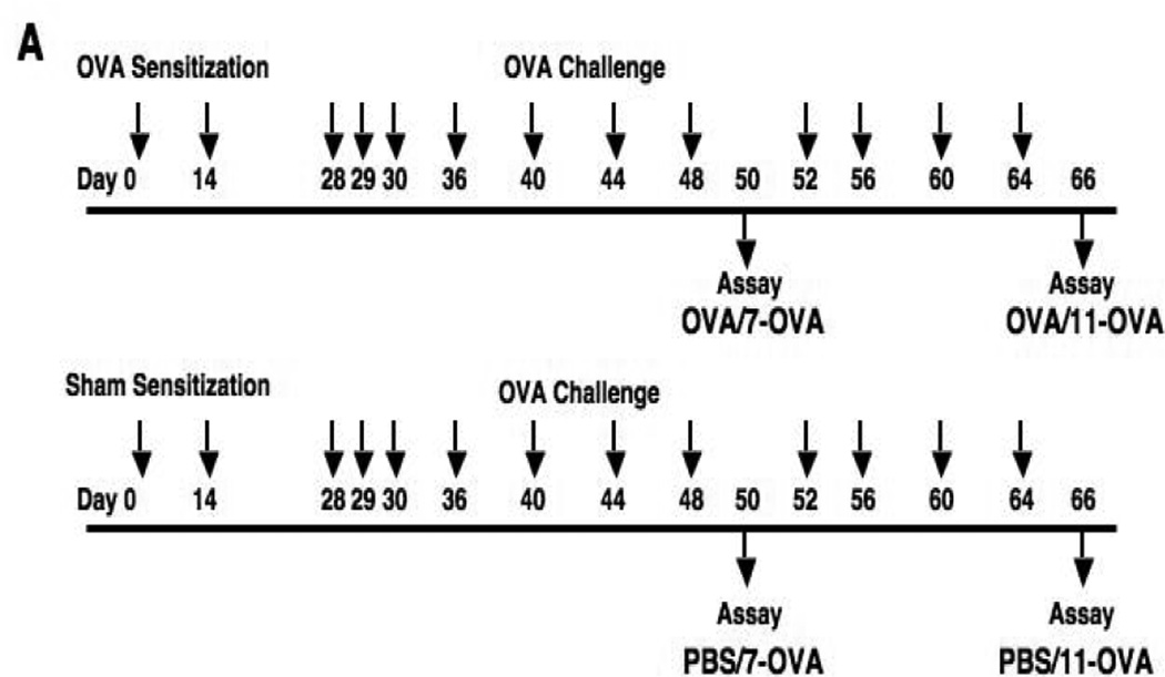
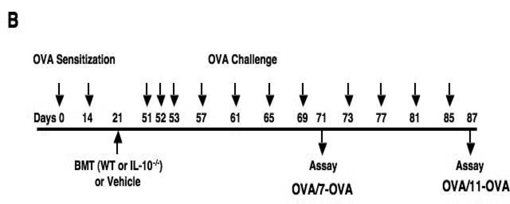
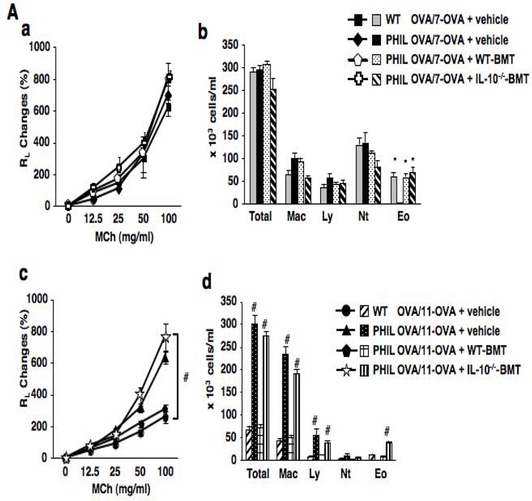
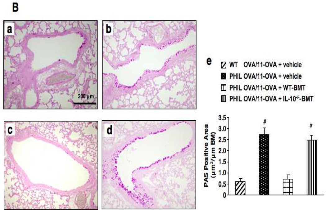
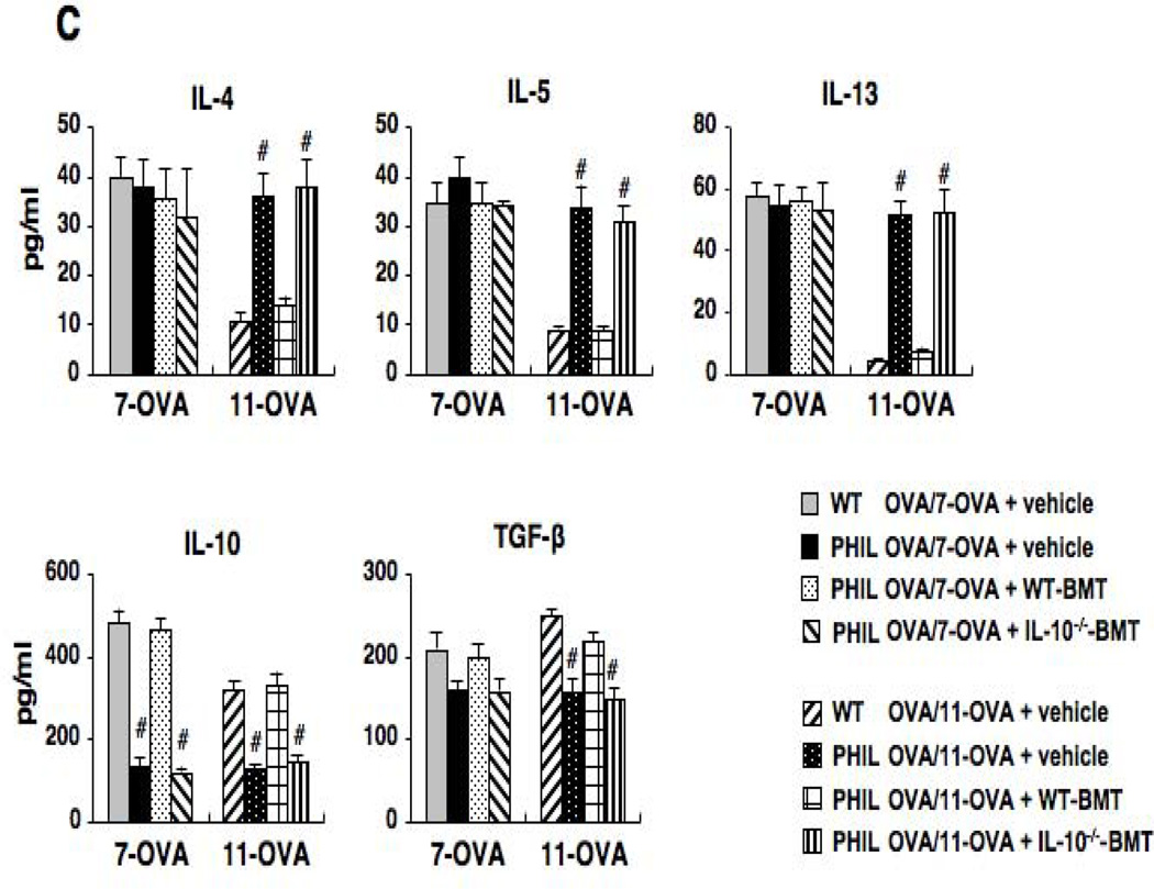
Methods: Eosinophil-deficient (PHIL) and wild-type (WT) littermates were sensitized and challenged to ovalbumin (OVA) 7 or 11 times. Airway inflammation, airway hyperresponsiveness (AHR) to inhaled methacholine, bronchoalveolar lavage (BAL) cytokine levels, and lung histology were monitored. Intracellular cytokine levels in BAL leukocytes were analyzed by flow cytometry. Groups of OVA-sensitized PHIL mice received bone marrow from WT or IL-10(-/-) donors 30 days before the OVA challenge.

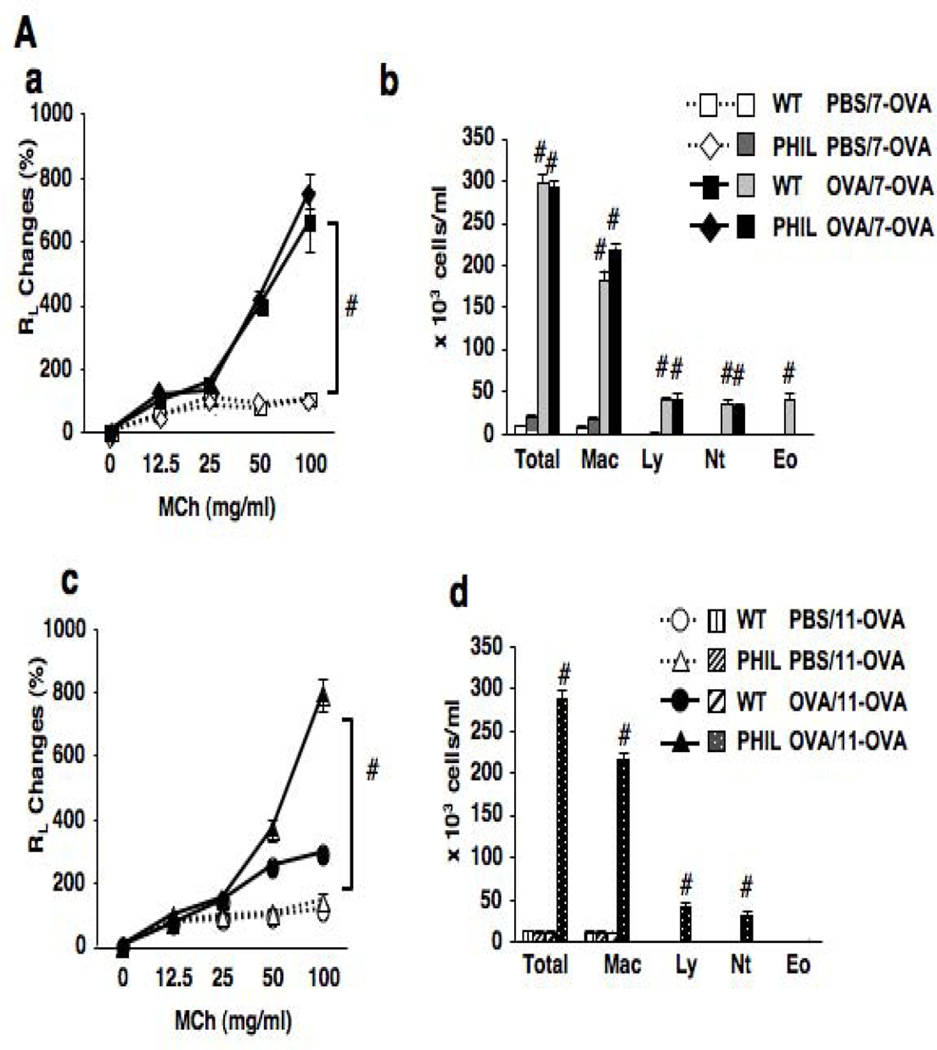
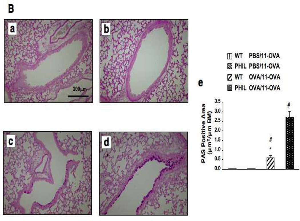
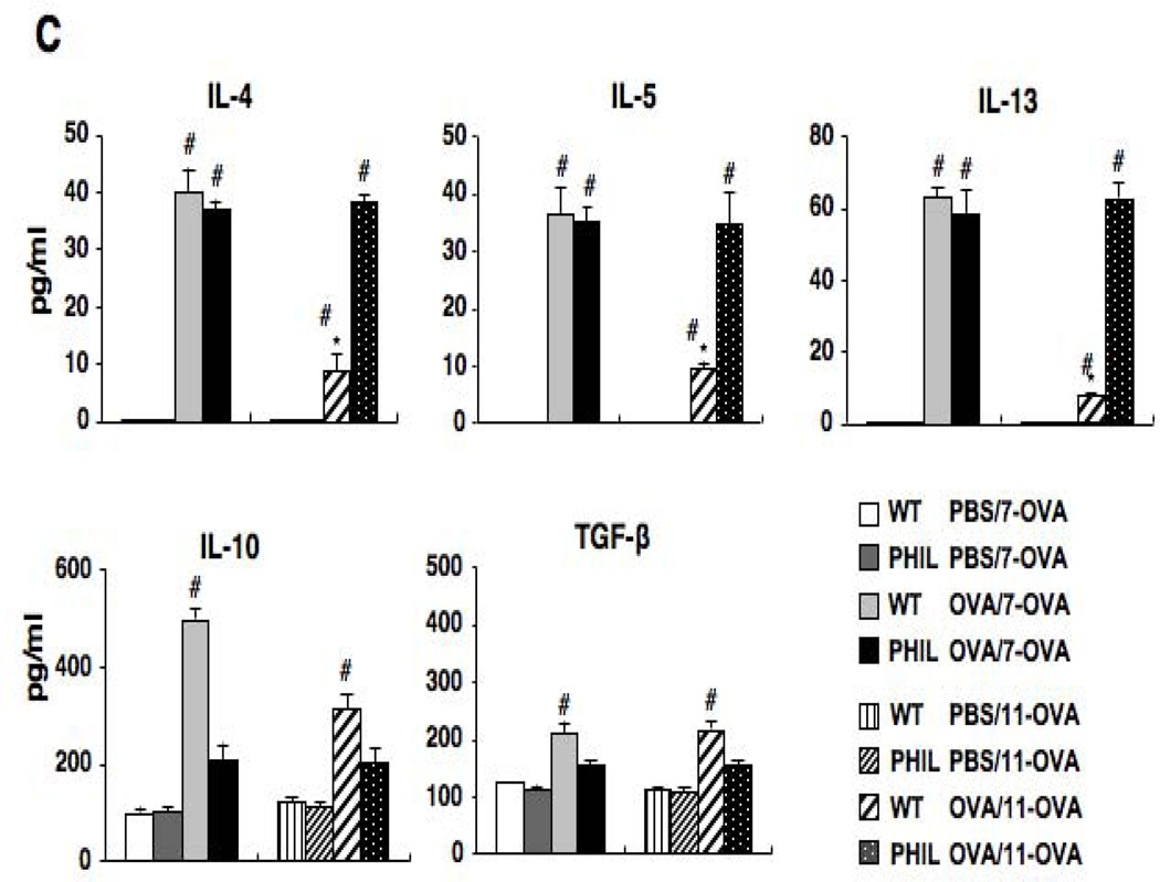
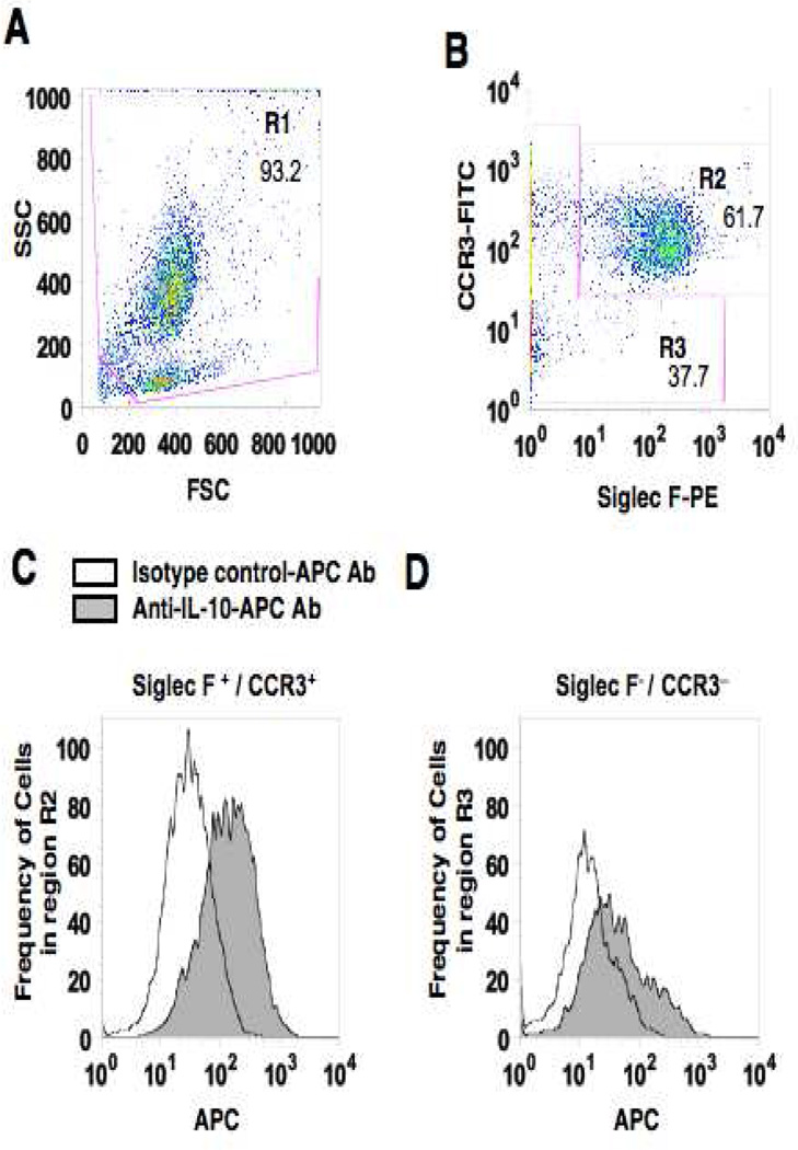
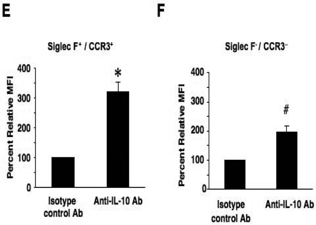
Results: PHIL and WT mice developed similar levels of AHR and numbers of leukocytes and cytokine levels in BAL fluid after OVA sensitization and 7 airway challenges; no eosinophils were detected in the PHIL mice. Unlike WT mice, sensitized PHIL mice maintained AHR, lung inflammation, and increased levels of IL-4, IL-5, and IL-13 in BAL fluid after 11 challenges whereas IL-10 and TGF-β levels were decreased. Restoration of eosinophil numbers after injection of bone marrow from WT but not IL-10-deficient mice restored levels of IL-10 and TGF-β in BAL fluid as well as suppressed AHR and inflammation. Intracellular staining of BAL leukocytes revealed the capacity of eosinophils to produce IL-10.

Conclusions: After repeated allergen challenge, eosinophils appeared not essential for the development of AHR and lung inflammation but contributed to the resolution of AHR and inflammation by producing IL-10.

Keywords: Eosinophils; IL-10; resolution of inflammation.


Figures

Figure 1
Figure 1
Experimental protocol for sensitization and challenge with OVA. (A) PHIL mice and WT littermates were sensitized to OVA on days 0 and 14 followed by aerosolized OVA challenges 7 times (OVA/OVA-7) or 11 times (OVA/OVA-11). Control mice received sham sensitization (PBS/OVA-7 and PBS/OVA-11). (B) Mice were sensitized with OVA in the same manner as described above. One week after the last sensitization, mice received bone marrow cells (5×10⁶) from WT or IL-10−/− mice. OVA challenges were initiated 30 days following bone marrow cell transfer. Controls received vehicle only.
Figure 1
Figure 1
Experimental protocol for sensitization and challenge with OVA. (A) PHIL mice and WT littermates were sensitized to OVA on days 0 and 14 followed by aerosolized OVA challenges 7 times (OVA/OVA-7) or 11 times (OVA/OVA-11). Control mice received sham sensitization (PBS/OVA-7 and PBS/OVA-11). (B) Mice were sensitized with OVA in the same manner as described above. One week after the last sensitization, mice received bone marrow cells (5×10⁶) from WT or IL-10−/− mice. OVA challenges were initiated 30 days following bone marrow cell transfer. Controls received vehicle only.
Figure 2
Figure 2
Changes in airway function and inflammation following repeated OVA challenges in WT and PHIL mice. (A) (a) Changes in lung resistance (RL) following 7 OVA challenges. (b) BAL cell composition. (c) Airway responsiveness following 11 OVA challenges. (d) BAL cell composition. n=6–9. #p<0.05 vs. PBS/OVA-7 or PBS/OVA-11 groups. (B) Goblet cell metaplasia. Representative photomicrographs of lung tissue from (a) WT mice following sham sensitization and 11 OVA challenges, (b) PHIL mice following sham sensitization and 11 OVA challenges, (c) WT mice following sensitization and 11 OVA challenges. (d) PHIL mice following sensitization and 11 OVA challenges. (e) Quantitation of PAS-positive areas along the airways. (C) BAL cytokine levels in WT and PHIL mice. BAL fluid samples were collected 48 hrs after 7 or 11 OVA challenges. n=9 in all experiments except panel B-e (n=6). Mac; macrophages, Ly; lymphocytes, Eo; eosinophils, Nt; neutrophils. #p<0.05 vs. PBS/OVA-7 and PBS/OVA-11. *p<0.05 vs. PHIL OVA/OVA-11.
Figure 2
Figure 2
Changes in airway function and inflammation following repeated OVA challenges in WT and PHIL mice. (A) (a) Changes in lung resistance (RL) following 7 OVA challenges. (b) BAL cell composition. (c) Airway responsiveness following 11 OVA challenges. (d) BAL cell composition. n=6–9. #p<0.05 vs. PBS/OVA-7 or PBS/OVA-11 groups. (B) Goblet cell metaplasia. Representative photomicrographs of lung tissue from (a) WT mice following sham sensitization and 11 OVA challenges, (b) PHIL mice following sham sensitization and 11 OVA challenges, (c) WT mice following sensitization and 11 OVA challenges. (d) PHIL mice following sensitization and 11 OVA challenges. (e) Quantitation of PAS-positive areas along the airways. (C) BAL cytokine levels in WT and PHIL mice. BAL fluid samples were collected 48 hrs after 7 or 11 OVA challenges. n=9 in all experiments except panel B-e (n=6). Mac; macrophages, Ly; lymphocytes, Eo; eosinophils, Nt; neutrophils. #p<0.05 vs. PBS/OVA-7 and PBS/OVA-11. *p<0.05 vs. PHIL OVA/OVA-11.
Figure 3
Figure 3
Consequences of eosinophil restoration following transfer of bone marrow cells. (A) (a) Lung resistance following bone marrow cell transfer and 7 OVA challenges. (b) BAL cell composition. (c) Lung resistance following bone marrow cell transfer and 11 OVA challenges. (d) BAL cell composition after 11 OVA challenges. (B) Goblet cell metaplasia. Representative photomicrographs in the lung tissue from (a) vehicle-treated WT mice after sensitization and 11 OVA challenges, (b) vehicle-treated PHIL mice after sensitization and 11 OVA challenges, (c) PHIL mice which received bone marrow cells from WT mice after sensitization and 11 OVA challenges, (d) PHIL mice which received bone marrow cells from IL-10−/− mice after sensitization and 11 OVA challenges, and (e) Quantitation of PAS-positive areas along the airways. (C) BAL fluid cytokine levels following bone marrow cell transfer and 11 OVA challenges. n=9 in all experiments except panel B-e (n=6). *p<0.05 vs. PHIL OVA/OVA-7+vehicle group, #p<0.05 vs. WT OVA/OVA-11+vehicle and PHIL OVA/OVA-11+ WT-BM.
Figure 3
Figure 3
Consequences of eosinophil restoration following transfer of bone marrow cells. (A) (a) Lung resistance following bone marrow cell transfer and 7 OVA challenges. (b) BAL cell composition. (c) Lung resistance following bone marrow cell transfer and 11 OVA challenges. (d) BAL cell composition after 11 OVA challenges. (B) Goblet cell metaplasia. Representative photomicrographs in the lung tissue from (a) vehicle-treated WT mice after sensitization and 11 OVA challenges, (b) vehicle-treated PHIL mice after sensitization and 11 OVA challenges, (c) PHIL mice which received bone marrow cells from WT mice after sensitization and 11 OVA challenges, (d) PHIL mice which received bone marrow cells from IL-10−/− mice after sensitization and 11 OVA challenges, and (e) Quantitation of PAS-positive areas along the airways. (C) BAL fluid cytokine levels following bone marrow cell transfer and 11 OVA challenges. n=9 in all experiments except panel B-e (n=6). *p<0.05 vs. PHIL OVA/OVA-7+vehicle group, #p<0.05 vs. WT OVA/OVA-11+vehicle and PHIL OVA/OVA-11+ WT-BM.
Figure 4
Figure 4
Intracellular IL-10 staining of airway eosinophils. BAL leukocytes from OVA sensitized and 7-challenged WT mice were activated with PMA/ionomysin and then stained with anti-mouse CCR3-FITC, anti-mouse Siglec-F-PE and anti-mouse IL-10-APC or APC-conjugated isotype control antibody. To identify the source of IL-10 in BAL fluid, live cells were first gated (region 1, R1) on forward scatter (FSC) and side scatter (SSC) (panel A). Cells in R1 were separated into Siglec-F- and CCR3-double-positive cells (R2) or negative cells (R3) (panel B). IL-10-positive cells in R2 or R3 were expressed as histograms (panel C and D) and the values for MFI were analyzed and expressed as the ratio to MFIs of isotype control staining in each sample (panels E and F). n=6. *p<0.001 and #p<0.01 vs. isotype control antibody group.
Figure 4
Figure 4
Intracellular IL-10 staining of airway eosinophils. BAL leukocytes from OVA sensitized and 7-challenged WT mice were activated with PMA/ionomysin and then stained with anti-mouse CCR3-FITC, anti-mouse Siglec-F-PE and anti-mouse IL-10-APC or APC-conjugated isotype control antibody. To identify the source of IL-10 in BAL fluid, live cells were first gated (region 1, R1) on forward scatter (FSC) and side scatter (SSC) (panel A). Cells in R1 were separated into Siglec-F- and CCR3-double-positive cells (R2) or negative cells (R3) (panel B). IL-10-positive cells in R2 or R3 were expressed as histograms (panel C and D) and the values for MFI were analyzed and expressed as the ratio to MFIs of isotype control staining in each sample (panels E and F). n=6. *p<0.001 and #p<0.01 vs. isotype control antibody group.

Comment in

References

    1. Bousquet J, Mantzouranis E, Cruz AA, Ait-Khaled N, Baena-Cagnani, Bleecker ER, et al. Uniform definition of asthma severity, control, and exacerbations: document presented for the World Health Organization Consultation on Severe Asthma. J Allergy Clin Immunol. 2010;126:926–938. - PubMed
    1. Zimmermann N, Hershey GK, Foster PS, Rothenberg ME. Chemokines in asthma: cooperative interaction between chemokines and IL-13. J Allergy Clin Immunol. 2003;111:227–242. quiz 243. - PubMed
    1. Wills-Karp M, Karp CL. Biomedicine. Eosinophils in asthma: remodeling a tangled tale. Science. 2004;305:1726–1729. - PubMed
    1. Kallinich T, Beier KC, Wahn U, Stock P, Hamelmann E. T-cell co-stimulatory molecules: their role in allergic immune reactions. Eur Respir J. 2007;29:1246–1255. - PubMed
    1. Rothenberg ME, Hogan SP. The eosinophil. Annu Rev Immunol. 2006;24:147–174. - PubMed

Publication types

MeSH terms