

Baseline effects of transcranial direct current stimulation on glutamatergic neurotransmission and large-scale network connectivity
- PMID: 25312829
- PMCID: PMC4358793
- DOI: 10.1016/j.brainres.2014.09.066
Baseline effects of transcranial direct current stimulation on glutamatergic neurotransmission and large-scale network connectivity
Abstract
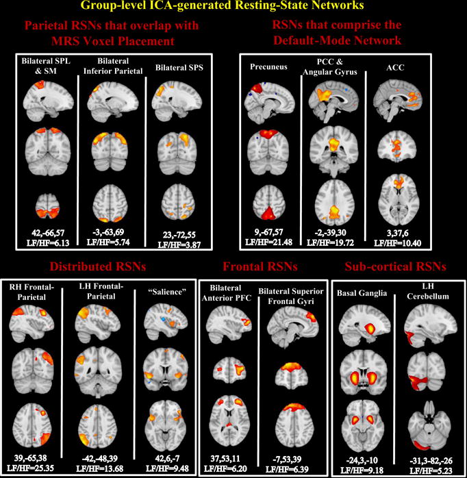
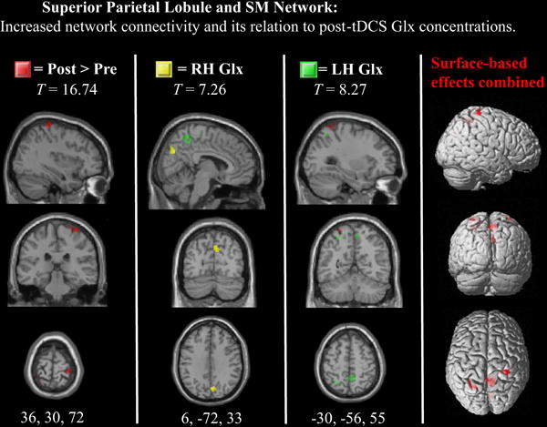
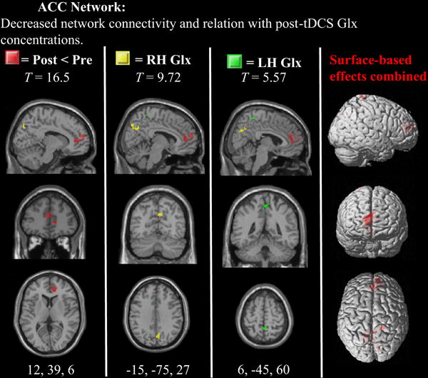
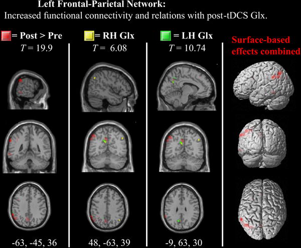
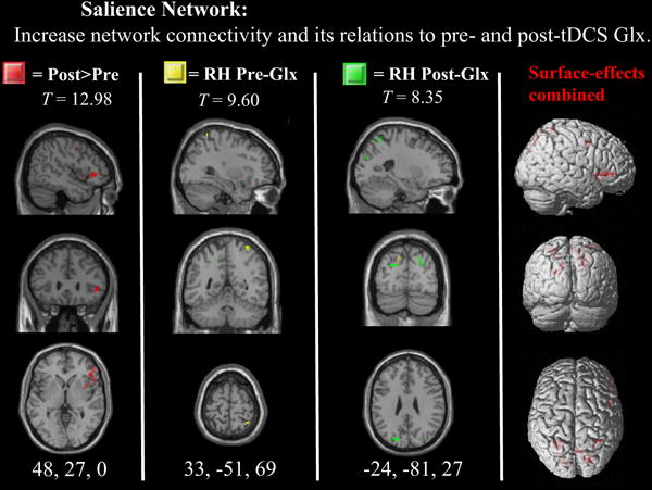
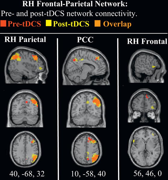
Transcranial direct current stimulation (tDCS) modulates glutamatergic neurotransmission and can be utilized as a novel treatment intervention for a multitude of populations. However, the exact mechanism by which tDCS modulates the brain׳s neural architecture, from the micro to macro scales, have yet to be investigated. Using a within-subjects design, resting-state functional magnetic resonance imaging (rs-fMRI) and proton magnetic resonance spectroscopy ((1)H MRS) were performed immediately before and after the administration of anodal tDCS over right parietal cortex. Group independent component analysis (ICA) was used to decompose fMRI scans into 75 brain networks, from which 12 resting-state networks were identified that had significant voxel-wise functional connectivity to anatomical regions of interest. (1)H MRS was used to obtain estimates of combined glutamate and glutamine (Glx) concentrations from bilateral intraparietal sulcus. Paired sample t-tests showed significantly increased Glx under the anodal electrode, but not in homologous regions of the contralateral hemisphere. Increases of within-network connectivity were observed within the superior parietal, inferior parietal, left frontal-parietal, salience and cerebellar intrinsic networks, and decreases in connectivity were observed in the anterior cingulate and the basal ganglia (p<0.05, FDR-corrected). Individual differences in Glx concentrations predicted network connectivity in most of these networks. The observed relationships between glutamatergic neurotransmission and network connectivity may be used to guide future tDCS protocols that aim to target and alter neuroplastic mechanisms in healthy individuals as well as those with psychiatric and neurologic disorders.
Keywords: Functional network connectivity (FNC); Glutamine–glutamate (Glx); Independent component analysis (ICA); Magnetic resonance spectroscopy (MRS); Parietal lobe; Resting-state functional magnetic resonance imaging (rs-fMRI); Transcranial direct current stimulation (tDCS).
Copyright © 2014 Elsevier B.V. All rights reserved.
Conflict of interest statement
Figures
Similar articles
-
Effects of bifrontal transcranial direct current stimulation on brain glutamate levels and resting state connectivity: multimodal MRI data for the cathodal stimulation site.Eur Arch Psychiatry Clin Neurosci. 2021 Feb;271(1):111-122. doi: 10.1007/s00406-020-01177-0. Epub 2020 Aug 2. Eur Arch Psychiatry Clin Neurosci. 2021. PMID: 32743758 Free PMC article. Clinical Trial.
-
Transcranial direct current stimulation reconstructs diminished thalamocortical connectivity during prolonged resting wakefulness: a resting-state fMRI pilot study.Brain Imaging Behav. 2020 Feb;14(1):278-288. doi: 10.1007/s11682-018-9979-9. Brain Imaging Behav. 2020. PMID: 30430411
-
Impact of network-targeted multichannel transcranial direct current stimulation on intrinsic and network-to-network functional connectivity.J Neurosci Res. 2020 Oct;98(10):1843-1856. doi: 10.1002/jnr.24690. Epub 2020 Jul 20. J Neurosci Res. 2020. PMID: 32686203 Free PMC article.
-
Glutamatergic neurometabolite levels in major depressive disorder: a systematic review and meta-analysis of proton magnetic resonance spectroscopy studies.Mol Psychiatry. 2019 Jul;24(7):952-964. doi: 10.1038/s41380-018-0252-9. Epub 2018 Oct 12. Mol Psychiatry. 2019. PMID: 30315224 Free PMC article.
-
Glutamatergic neurotransmission in schizophrenia: A systematic review and quantitative synthesis of proton magnetic resonance spectroscopy studies across schizophrenia spectrum disorders.Aust N Z J Psychiatry. 2024 Nov;58(11):930-951. doi: 10.1177/00048674241254216. Epub 2024 May 29. Aust N Z J Psychiatry. 2024. PMID: 38812258 Free PMC article.
Cited by
-
Reductions in GABA following a tDCS-language intervention for primary progressive aphasia.Neurobiol Aging. 2019 Jul;79:75-82. doi: 10.1016/j.neurobiolaging.2019.03.011. Epub 2019 Mar 27. Neurobiol Aging. 2019. PMID: 31029018 Free PMC article.
-
Dopamine Alters the Effect of Brain Stimulation on Decision-Making.J Neurosci. 2023 Oct 11;43(41):6909-6919. doi: 10.1523/JNEUROSCI.1140-23.2023. Epub 2023 Aug 30. J Neurosci. 2023. PMID: 37648451 Free PMC article.
-
Virtual reality for the treatment of neuropathic pain in people with spinal cord injuries: A scoping review.J Spinal Cord Med. 2021 Jan;44(1):8-18. doi: 10.1080/10790268.2019.1575554. Epub 2019 Feb 1. J Spinal Cord Med. 2021. PMID: 30707649 Free PMC article.
-
Understanding and targeting repetitive behaviors and restricted interests in autism spectrum disorder via high-definition transcranial direct current stimulation: a study-protocol.BMC Psychiatry. 2025 Feb 25;25(1):170. doi: 10.1186/s12888-025-06506-y. BMC Psychiatry. 2025. PMID: 40001028 Free PMC article.
-
Effects of transcranial direct current stimulation over right posterior parietal cortex on attention function in healthy young adults.Eur J Neurosci. 2019 Jun;49(12):1623-1631. doi: 10.1111/ejn.14349. Epub 2019 Feb 13. Eur J Neurosci. 2019. PMID: 30667550 Free PMC article.
References
-
- Corbetta M, Akbudak E, Conturo TE, Snyder AZ, Ollinger JM, Drury HA, Shulman GL. A Common Network of Functional Areas for Attention and Eye Movements. Neuron. 1998;21(4):761–773. - PubMed
Publication types
MeSH terms
Substances
Grants and funding
LinkOut - more resources
Full Text Sources
Other Literature Sources
Miscellaneous
