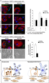
Decreased expression of KGF/FGF7 and its receptor in pathological hypopigmentation
- PMID: 25313018
- PMCID: PMC4302659
- DOI: 10.1111/jcmm.12411
Decreased expression of KGF/FGF7 and its receptor in pathological hypopigmentation
Figures


Similar articles
-
Melanosome transfer promoted by keratinocyte growth factor in light and dark skin-derived keratinocytes.J Invest Dermatol. 2008 Mar;128(3):558-67. doi: 10.1038/sj.jid.5701063. Epub 2007 Sep 20. J Invest Dermatol. 2008. PMID: 17882267
-
Expression and signaling of the tyrosine kinase FGFR2b/KGFR regulates phagocytosis and melanosome uptake in human keratinocytes.FASEB J. 2011 Jan;25(1):170-81. doi: 10.1096/fj.10-162156. Epub 2010 Sep 15. FASEB J. 2011. PMID: 20844240
-
[Expression of keratinocyte growth factor and keratinocyte growth factor receptor and its impact on Hela cells].Sichuan Da Xue Xue Bao Yi Xue Ban. 2007 Jun;38(3):437-9. Sichuan Da Xue Xue Bao Yi Xue Ban. 2007. PMID: 17593825 Chinese.
-
An overview on keratinocyte growth factor: from the molecular properties to clinical applications.Protein Pept Lett. 2014 Mar;21(3):306-17. doi: 10.2174/09298665113206660115. Protein Pept Lett. 2014. PMID: 24188496 Review.
-
Keratinocyte growth factor expression and activity in cancer: implications for use in patients with solid tumors.J Natl Cancer Inst. 2006 Jun 21;98(12):812-24. doi: 10.1093/jnci/djj228. J Natl Cancer Inst. 2006. PMID: 16788155 Review.
Cited by
-
Tracking footprints of artificial and natural selection signatures in breeding and non-breeding cats.Sci Rep. 2022 Oct 27;12(1):18061. doi: 10.1038/s41598-022-22155-7. Sci Rep. 2022. PMID: 36302822 Free PMC article.
-
Skin Pigmentation and Pigmentary Disorders: Focus on Epidermal/Dermal Cross-Talk.Ann Dermatol. 2016 Jun;28(3):279-89. doi: 10.5021/ad.2016.28.3.279. Epub 2016 May 25. Ann Dermatol. 2016. PMID: 27274625 Free PMC article. Review.
-
Vitiligo and Autoimmune Thyroid Disorders.Front Endocrinol (Lausanne). 2017 Oct 27;8:290. doi: 10.3389/fendo.2017.00290. eCollection 2017. Front Endocrinol (Lausanne). 2017. PMID: 29163360 Free PMC article. Review.
-
Emerging Roles of Dermal Fibroblasts in Hyperpigmentation and Hypopigmentation: A Review.J Cosmet Dermatol. 2025 Jan;24(1):e16790. doi: 10.1111/jocd.16790. J Cosmet Dermatol. 2025. PMID: 39780507 Free PMC article. Review.
-
Fibroblast: A Novel Target for Autoimmune and Inflammatory Skin Diseases Therapeutics.Clin Rev Allergy Immunol. 2024 Jun;66(3):274-293. doi: 10.1007/s12016-024-08997-1. Epub 2024 Jun 28. Clin Rev Allergy Immunol. 2024. PMID: 38940997 Review.
References
-
- Seiberg M, Paine C, Sharlow E, et al. The protease-activated receptor-2 regulates pigmentation via keratinocyte-melanocyte interactions. Exp Cell Res. 2000;254:25–32. - PubMed
-
- Cardinali G, Ceccarelli S, Kovacs D, et al. Keratinocyte growth factor promotes melanosome transfer to keratinocytes. J Invest Dermatol. 2005;125:1190–9. - PubMed
-
- Van Den Bossche K, Naeyaert JM, Lambert J. The quest for the mechanism of melanin transfer. Traffic. 2006;7:1–10. - PubMed
-
- Cardinali G, Bolasco G, Aspite N, et al. Melanosome transfer promoted by keratinocyte growth factor in light and dark skin-derived keratinocytes. J Invest Dermatol. 2008;128:558–67. - PubMed
-
- Belleudi F, Purpura V, Scrofani C, et al. Expression and signaling of the tyrosine kinase FGFR2b/KGFR regulates phagocytosis and melanosome uptake in human keratinocytes. FASEB J. 2011;25:170–81. - PubMed
Publication types
MeSH terms
Substances
LinkOut - more resources
Full Text Sources
Other Literature Sources

