. 2014 Oct 28;111(43):15379-84.
doi: 10.1073/pnas.1410356111. Epub 2014 Oct 13.

Sequence selectivity of macrolide-induced translational attenuation

Affiliations

Sequence selectivity of macrolide-induced translational attenuation

Amber R Davis et al. Proc Natl Acad Sci U S A. .

Abstract

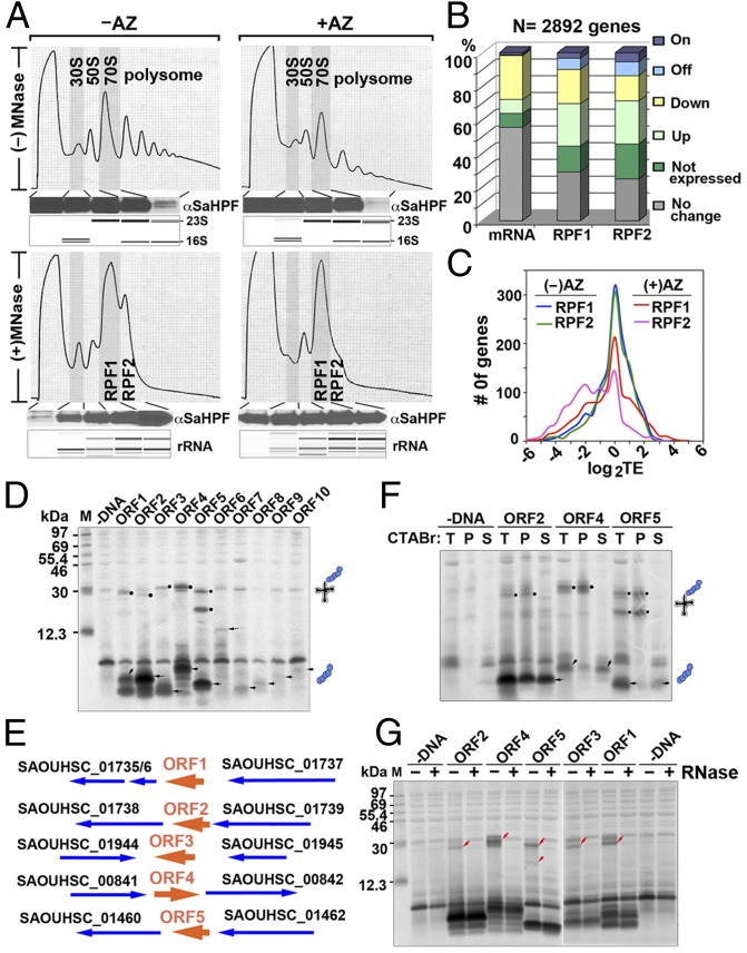
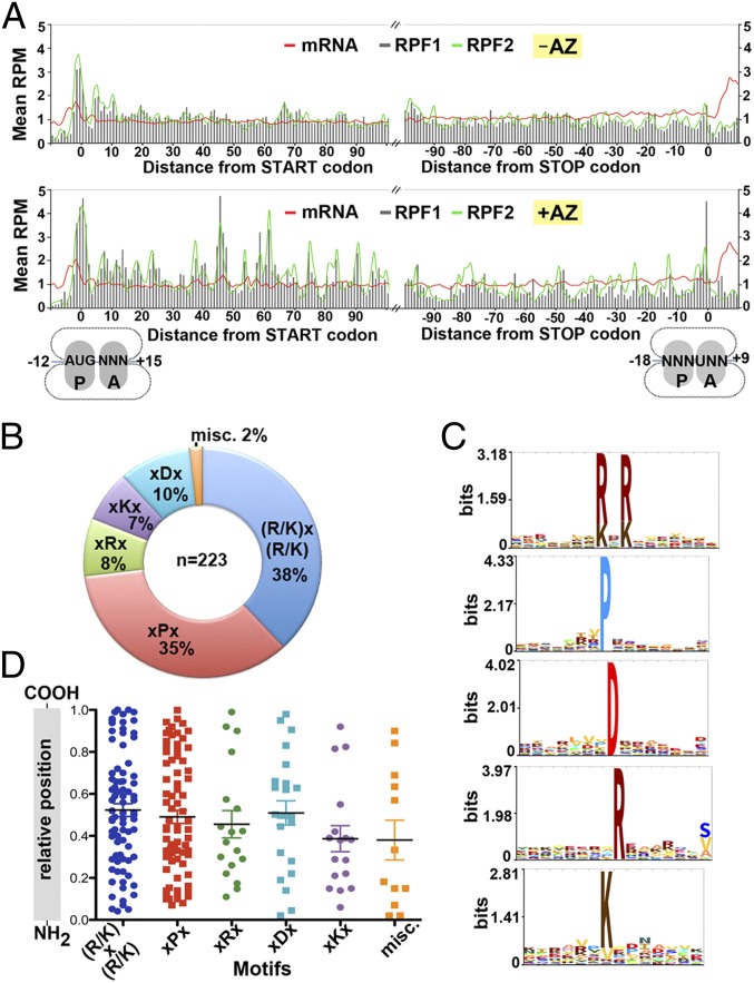
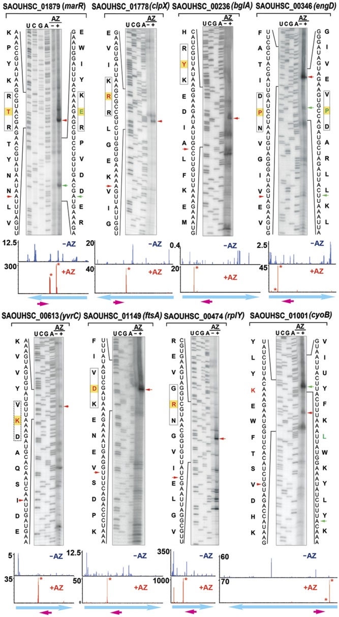
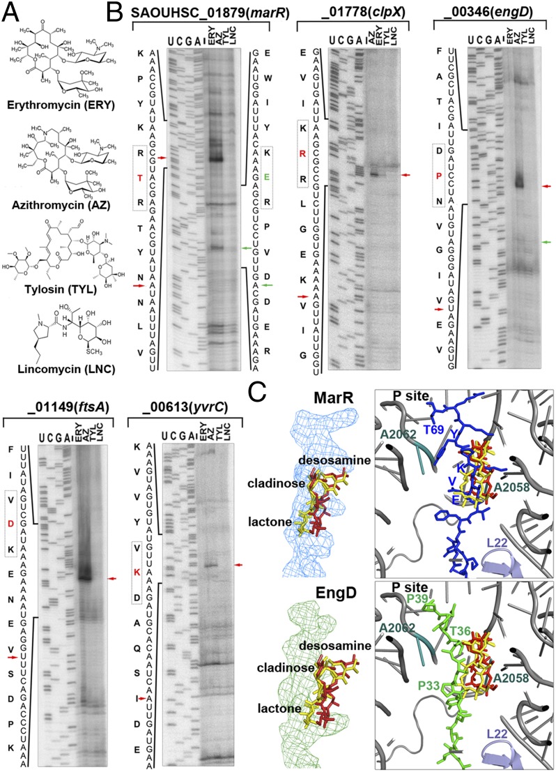
The prevailing "plug-in-the-bottle" model suggests that macrolide antibiotics inhibit translation by binding inside the ribosome tunnel and indiscriminately arresting the elongation of every nascent polypeptide after the synthesis of six to eight amino acids. To test this model, we performed a genome-wide analysis of translation in azithromycin-treated Staphylococcus aureus. In contrast to earlier predictions, we found that the macrolide does not preferentially induce ribosome stalling near the 5' end of mRNAs, but rather acts at specific stalling sites that are scattered throughout the entire coding region. These sites are highly enriched in prolines and charged residues and are strikingly similar to other ligand-independent ribosome stalling motifs. Interestingly, the addition of structurally related macrolides had dramatically different effects on stalling efficiency. Our data suggest that ribosome stalling can occur at a surprisingly large number of low-complexity motifs in a fashion that depends only on a few arrest-inducing residues and the presence of a small molecule inducer.

Keywords: Staphylococcus aureus; antibiotic; ribosome stalling.


Conflict of interest statement

The authors declare no conflict of interest.

Figures

Fig. 1.
Fig. 1.
Analysis of RPFs and identification of previously unidentified ORFs. (A) Sucrose gradient profiles of S. aureus ribosomes digested and undigested by MNase with or without AZ treatment. The y axis corresponds to the absorbance at 254 nm of the ribosome particles separated on a 10–55% sucrose gradient. The association of SaHPF to the 100S ribosome, which constituted >85% of the RPF2 (Fig. S1F), was detected via immunoblotting. (Lower) Bioanalyzer electropherograms of the RNA species are shown as controls. (B) Global effects of AZ on the transcriptome and the translatome. Differential gene expression is defined as ≥twofold changes (<0.05 false discovery rate) in the RPKM (reads per kilobase of transcript per million mapped reads) ratios between the AZ-treated to -untreated samples. “On” and “Off” are defined as genes that are transcriptionally or translationally inactive (zero read count) in one sample. (C) Translation efficiency (TE) was moderately decreased after AZ exposure. The efficiency was calculated as the log2 ratios of the RPFs to the mRNA fragments that were measured in RPKM. (D) Cell-free coupled transcription–translation of representative short ORFs. Arrowheads indicate the protein products, and solid circles denote stalled peptidyl-tRNAs. (E) Genetic organization of arrest peptide-encoded ORFs (in orange). Genes are depicted by thick arrows, which point in the direction of transcription. ORF1–ORF5 undergo translation arrest without AZ. (F) CTABr precipitation of peptidyl-tRNAs that remain tethered to the stalled ribosomes. Full-length proteins were fractionated into the CTABr supernatant. T, total; P, pellet; S, supernatant. (G) The RNase A susceptibility of translation-arrested peptidyl-tRNAs is indicated by the disappearance of the ∼20- to 30-kDa bands (red arrowheads).
Fig. 2.
Fig. 2.
AZ-specific ribosome stalling sites are scattered along ORFs and are enriched in proline and charged residues. (A) Ribosome density as a function of position. Metagene analysis of read densities in the AZ-treated (Lower) and untreated (Upper) samples. The normalized RPM correspond to the average ribosome density across the most abundantly translated ORFs (>50 counts), which were aligned relative to the start and stop codons. Total mRNA and RPF2 are shown with red and green lines, respectively. RPF1 is depicted by gray bars. (B) The fraction of stalling sequences falling into each indicated class. (C) Hidden Markov model logos showing the frequencies of specific residues at a given position in each motif group. Sequences of miscellaneous class are too variable to build the logo. (D) The relative location of the stalling motifs within their full-length proteins. Error bars indicate mean.
Fig. 3.
Fig. 3.
In vitro mapping of AZ-specific ribosome stalling sites by toe-printing analysis. Cell-free translation was programmed with the respective DNA templates in the presence and absence of AZ, followed by primer extension. Tripeptide stalling sequences are boxed. The residues labeled in red and green are positioned in the P site of the stalled ribosome, which are located 16–17 nt upstream of the blocked reverse transcriptase. Red and green arrowheads mark the primary and secondary toe-print signals. The ribosome density maps corresponding to the genes are shown on the bottom panels. The y axis indicates RPM-normalized ribosome density. The magenta arrows indicate the location of the reverse primer. The stalling sites are labeled with an asterisk. Weaker density peaks might not be visible owing to the scale difference.
Fig. 4.
Fig. 4.
Distinct dispositions of MarR and EngD peptides inside the tunnel affect stalling efficiency. (A) Chemical structures of macrolides (ERY, AZ, and tylosin) and lincomycin. (B) Toe-printing analysis of representative stalling sequences in the presence of different antibiotics. Labeling schemes are identical to those of Fig. 3. (C) Structural modeling of the MarR and EngD stalling peptides inside the antibiotic-bound tunnel. The relative locations of previously reported ribosome stalling “sensors” (L22, A2062, and A2058) to the superimposed ERY (red) and AZ (yellow) molecules are indicated.

References

    1. Nissen P, Hansen J, Ban N, Moore PB, Steitz TA. The structural basis of ribosome activity in peptide bond synthesis. Science. 2000;289(5481):920–930. - PubMed
    1. Bhushan S, et al. alpha-Helical nascent polypeptide chains visualized within distinct regions of the ribosomal exit tunnel. Nat Struct Mol Biol. 2010;17(3):313–317. - PubMed
    1. Lu J, Deutsch C. Secondary structure formation of a transmembrane segment in Kv channels. Biochemistry. 2005;44(23):8230–8243. - PubMed
    1. Woolhead CA, Johnson AE, Bernstein HD. Translation arrest requires two-way communication between a nascent polypeptide and the ribosome. Mol Cell. 2006;22(5):587–598. - PubMed
    1. Ito K, Chiba S. Arrest peptides: Cis-acting modulators of translation. Annu Rev Biochem. 2013;82:171–202. - PubMed

Publication types

MeSH terms

Associated data