. 2014 Oct 28;111(43):E4606-14.
doi: 10.1073/pnas.1407020111. Epub 2014 Oct 13.

Capsid expansion mechanism of bacteriophage T7 revealed by multistate atomic models derived from cryo-EM reconstructions

Affiliations

Capsid expansion mechanism of bacteriophage T7 revealed by multistate atomic models derived from cryo-EM reconstructions

Fei Guo et al. Proc Natl Acad Sci U S A. .

Abstract

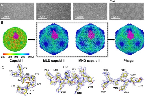
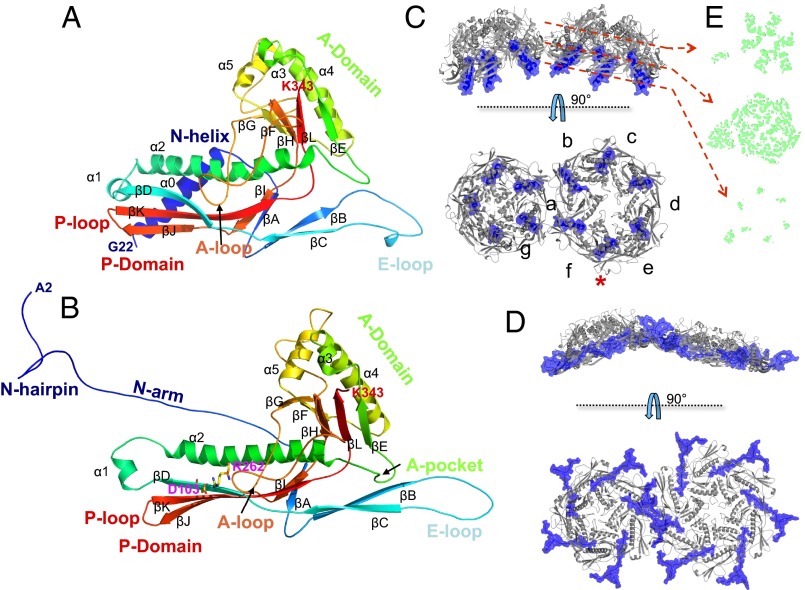
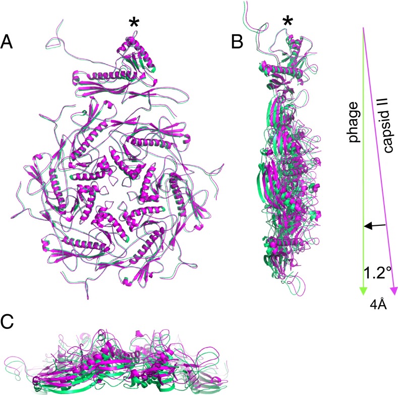
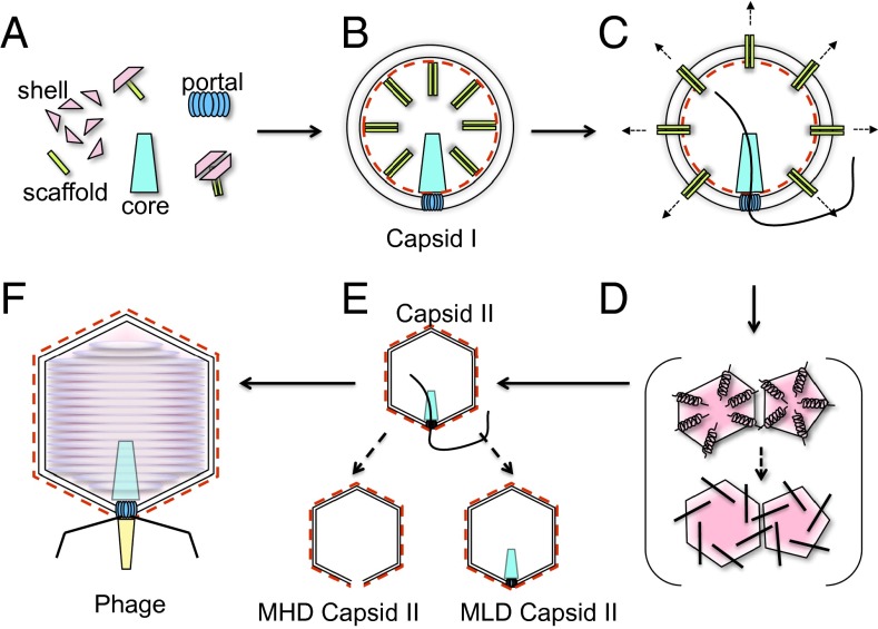
Many dsDNA viruses first assemble a DNA-free procapsid, using a scaffolding protein-dependent process. The procapsid, then, undergoes dramatic conformational maturation while packaging DNA. For bacteriophage T7 we report the following four single-particle cryo-EM 3D reconstructions and the derived atomic models: procapsid (4.6-Å resolution), an early-stage DNA packaging intermediate (3.5 Å), a later-stage packaging intermediate (6.6 Å), and the final infectious phage (3.6 Å). In the procapsid, the N terminus of the major capsid protein, gp10, has a six-turn helix at the inner surface of the shell, where each skewed hexamer of gp10 interacts with two scaffolding proteins. With the exit of scaffolding proteins during maturation the gp10 N-terminal helix unfolds and swings through the capsid shell to the outer surface. The refolded N-terminal region has a hairpin that forms a novel noncovalent, joint-like, intercapsomeric interaction with a pocket formed during shell expansion. These large conformational changes also result in a new noncovalent, intracapsomeric topological linking. Both interactions further stabilize the capsids by interlocking all pentameric and hexameric capsomeres in both DNA packaging intermediate and phage. Although the final phage shell has nearly identical structure to the shell of the DNA-free intermediate, surprisingly we found that the icosahedral faces of the phage are slightly (∼4 Å) contracted relative to the faces of the intermediate, despite the internal pressure from the densely packaged DNA genome. These structures provide a basis for understanding the capsid maturation process during DNA packaging that is essential for large numbers of dsDNA viruses.

Keywords: DNA packaging intermediates; bacteriophage T7 maturation; noncovalent topological linking; procapsid; single-particle cryo-EM.


Conflict of interest statement

The authors declare no conflict of interest.

Figures

Fig. 1.
Fig. 1.
Single-particle 3D reconstructions of bacteriophage T7. (A) Cryo-EM images of capsid I, MLD capsid II, MHD capsid II, and phage. (B) Threefold surface view of corresponding 3D reconstructions. The surfaces are colored by radius. One T = 7L asymmetric unit is highlighted in magenta. (C) Electron densities of three representative regions (a loop, an α-helix, and a β-strand) of MLD capsid II structure.
Fig. 2.
Fig. 2.
Atomic models of major capsid protein gp10A. (A) Subunit g in capsid I. (B) Subunit d in MLD capsid II. The models are rainbow-colored from N (blue) to C (red) terminus. A salt bridge formed by Asp-103 and Arg-262 is shown as sticks in B. A and B are viewed from the capsid outer surface, similar to the orientation of the bottom panel in C and D. (C and D) Arrangement of subunits in a pentamer and a hexamer of the (C) capsid I shell and (D) MLD II capsid II shell in two orthogonal views (side view in top panel and top view in bottom panel). The N-helix (C) and N-arm (D) regions are colored in blue. The red asterisk indicates the P-domain/E-loop interaction of subunits e and f. (E) The section views of the asymmetric unit density (green) of capsid I. The locations of the sections, which have variable degrees of skewing, are indicated by red dashed arrows.
Fig. 3.
Fig. 3.
Interactions of scaffolding protein gp9 with major capsid protein gp10 on the inner surface of capsid I. (A) The inner surface is radially colored. The scaffolding protein, gp9, densities (blue) are found on hexamers, but not on pentamers. Scaffolding protein in the boxed region is magnified in C. The electron densities were low-pass-filtered to 7 Å for cleaner view. (B) Zoom-in view of the N-helix region. The N-helix model (sticks) is superimposed in the electron density (wire frames). (C) Zoom-in view of the electron densities of scaffolding protein HTH region found on gp10 subunit f. (D) Model of gp10 and the gp9 HTH region. The gp10 is the same subunit, f, as shown in B.
Fig. 4.
Fig. 4.
Novel intracapsomere topological linking and intercapsomere joint in capsid II and phage. (A) Outer surface view of a capsid region slightly larger than an icosahedral face. Three hexamers are highlighted in different colors (red, blue, and yellow). (B) Magnified view of two neighboring hexamers with two interactions indicated by boxes. The first interaction is the intercapsomere joint interaction (magenta box) between the N-hairpin (red) and the A-pocket (blue). The E-loop, βD-strand, and A-loop regions of the hexamers are also colored to highlight the second interaction. The second interaction is intracapsomere nonconvalent topological linking (orange box). (C) Zoom-in view of the first interaction, that is, between the A-pocket (K151, Y152, N153, I156, and E157 shown as stick) and the N-hairpin (Q14, G15, K16, G17, V18, and V19 shown as stick) around twofold and pseudotwofold axes. Electron densities of this region are shown as wire frames. (D) Zoom-in view of the second interaction, which is formed by encircling of an E-loop (red) by a loop formed via a salt bridge (stick and wire frame) between R262 in the A-loop and D103 in the βD-strand of a neighboring subunit.
Fig. 5.
Fig. 5.
Comparison of asymmetric unit positions in the MLD capsid II and mature phage shells. The top view (A) and the side views (B and C) of MLD capsid II and phage asymmetric units are superimposed. MLD capsid II and phage are colored magenta and green, respectively. The asymmetric unit of MLD capsid II undergoes ∼1.2° inward tilt around the pivot point (marked by asterisk) to reach the final position in phage state.
Fig. 6.
Fig. 6.
Proposed assembly and maturation pathway of bacteriophage T7. (A) Capsid proteins and scaffolding proteins are expressed, along with other components. Three capsid proteins and one scaffolding protein form a building block, assumed to dimerize as a skewed hexameric capsomere. (B) These building blocks assemble on a partial capsid until the icosahedral cage is completed. The N-helix (red dashed line) of capsid protein is hidden inside the shell. (C) DNA packaging starts from the procapsid. Scaffolding proteins are expelled from the procapsid via the center openings of capsid protein hexamers to initiate expansion. (D) Having lost interaction with scaffolding proteins, capsid proteins undergo conformational changes that result in symmetric hexamers that are further stabilized by the newly formed intracapsomere, noncovalent, topological linking. Major conformational change occurs at the N terminus, where the N-helix at the inner surface unfolds, swings through the capsid shell, and becomes exposed on outer surface. The new N-terminal conformation results in joint-like strong interactions between neighboring capsomeres. (E) DNA is packaged in capsid II. Some are productive, but incomplete, and yield capsids (MLD capsid II) that lose partially packaged DNA during cell lysis and/or sample purification. Some of the capsid II particles at later packaging states would also lose portal and core stack proteins and yield MHD capsid II. (F) DNA packaging is completed with tail attachment, generating a mature phage particle.

Similar articles

Cited by

References

    1. Chen DH, et al. Structural basis for scaffolding-mediated assembly and maturation of a dsDNA virus. Proc Natl Acad Sci USA. 2011;108(4):1355–1360. - PMC - PubMed
    1. Ionel A, et al. Molecular rearrangements involved in the capsid shell maturation of bacteriophage T7. J Biol Chem. 2011;286(1):234–242. - PMC - PubMed
    1. Jiang W, et al. Coat protein fold and maturation transition of bacteriophage P22 seen at subnanometer resolutions. Nat Struct Biol. 2003;10(2):131–135. - PubMed
    1. Cerritelli ME, Conway JF, Cheng N, Trus BL, Steven AC. Molecular mechanisms in bacteriophage T7 procapsid assembly, maturation, and DNA containment. Adv Protein Chem. 2003;64:301–323. - PubMed
    1. Conway JF, et al. Virus maturation involving large subunit rotations and local refolding. Science. 2001;292(5517):744–748. - PubMed

Publication types

Substances