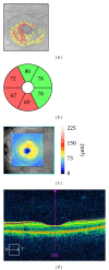
Comparative diagnostic accuracy of ganglion cell-inner plexiform and retinal nerve fiber layer thickness measures by Cirrus and Spectralis optical coherence tomography in relapsing-remitting multiple sclerosis
- PMID: 25313352
- PMCID: PMC4182893
- DOI: 10.1155/2014/128517
Comparative diagnostic accuracy of ganglion cell-inner plexiform and retinal nerve fiber layer thickness measures by Cirrus and Spectralis optical coherence tomography in relapsing-remitting multiple sclerosis
Abstract
Objective: To estimate sensitivity and specificity of several optical coherence tomography (OCT) measurements for detecting retinal thickness changes in patients with relapsing-remitting multiple sclerosis (RRMS), such as macular ganglion cell-inner plexiform layer (GCIPL) thickness measured with Cirrus (OCT) and peripapillary retinal nerve fiber layer (pRNFL) thickness measured with Cirrus and Spectralis OCT.
Methods: Seventy patients (140 eyes) with RRMS and seventy matched healthy subjects underwent pRNFL and GCIPL thickness analysis using Cirrus OCT and pRNFL using Spectralis OCT. A prospective, cross-sectional evaluation of sensitivities and specificities was performed using latent class analysis due to the absence of a gold standard.
Results: GCIPL measures had higher sensitivity and specificity than temporal pRNFL measures obtained with both OCT devices. Average GCIPL thickness was significantly more sensitive than temporal pRNFL by Cirrus (96.34% versus 58.41%) and minimum GCIPL thickness was significantly more sensitive than temporal pRNFL by Spectralis (96.41% versus 69.69%). Generalised estimating equation analysis revealed that age (P = 0.030), optic neuritis antecedent (P = 0.001), and disease duration (P = 0.002) were significantly associated with abnormal results in average GCIPL thickness.
Conclusion: Average and minimum GCIPL measurements had significantly better sensitivity to detect retinal thickness changes in RRMS than temporal pRNFL thickness measured by Cirrus and Spectralis OCT, respectively.
Figures


References
-
- Ojeda E, Díaz-Cortes D, Rosales D, Duarte-Rey C, Anaya J-M, Rojas-Villarraga A. Prevalence and clinical features of multiple sclerosis in Latin America. Clinical Neurology and Neurosurgery. 2013;115(4):381–387. - PubMed
-
- Ikuta F, Zimmerman HM. Distribution of plaques in seventy autopsy cases of multiple sclerosis in the United States. Neurology. 1976;26(6) part 2:26–28. - PubMed
-
- Rebolleda G, González-López JJ, Muñoz-Negrete FJ, Oblanca N, Costa-Frossard L, Álvarez-Cermeño JC. Color-code agreement among stratus, cirrus, and spectralis optical coherence tomography in relapsing-remitting multiple sclerosis with and without prior optic neuritis. The American Journal of Ophthalmology. 2013;155(5):890–897. - PubMed
Publication types
MeSH terms
LinkOut - more resources
Full Text Sources
Other Literature Sources

