. 2015 Jan 1;197(1):29-39.
doi: 10.1128/JB.02083-14. Epub 2014 Oct 13.

Rex (encoded by DVU_0916) in Desulfovibrio vulgaris Hildenborough is a repressor of sulfate adenylyl transferase and is regulated by NADH

Affiliations

Rex (encoded by DVU_0916) in Desulfovibrio vulgaris Hildenborough is a repressor of sulfate adenylyl transferase and is regulated by NADH

G A Christensen et al. J Bacteriol. .

Abstract

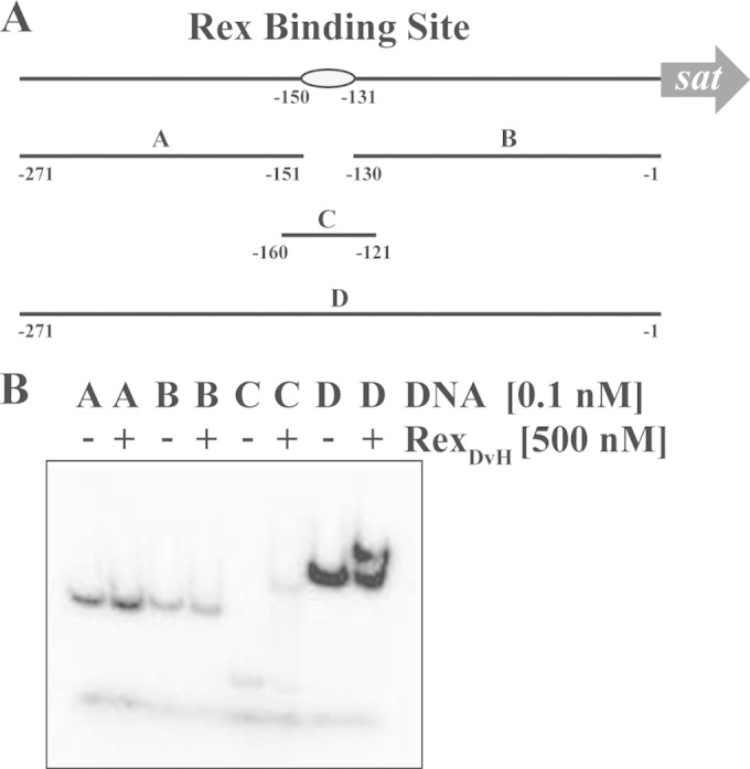
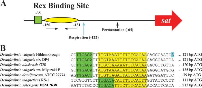
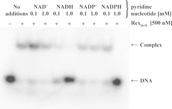
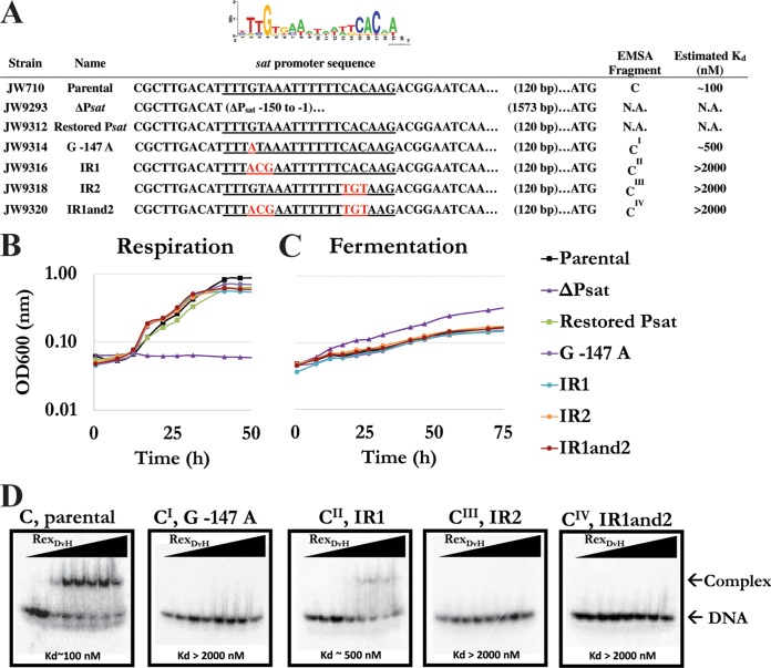
Although the enzymes for dissimilatory sulfate reduction by microbes have been studied, the mechanisms for transcriptional regulation of the encoding genes remain unknown. In a number of bacteria the transcriptional regulator Rex has been shown to play a key role as a repressor of genes producing proteins involved in energy conversion. In the model sulfate-reducing microbe Desulfovibrio vulgaris Hildenborough, the gene DVU_0916 was observed to resemble other known Rex proteins. Therefore, the DVU_0916 protein has been predicted to be a transcriptional repressor of genes encoding proteins that function in the process of sulfate reduction in D. vulgaris Hildenborough. Examination of the deduced DVU_0916 protein identified two domains, one a winged helix DNA-binding domain common for transcription factors, and the other a Rossman fold that could potentially interact with pyridine nucleotides. A deletion of the putative rex gene was made in D. vulgaris Hildenborough, and transcript expression studies of sat, encoding sulfate adenylyl transferase, showed increased levels in the D. vulgaris Hildenborough Rex (RexDvH) mutant relative to the parental strain. The RexDvH-binding site upstream of sat was identified, confirming RexDvH to be a repressor of sat. We established in vitro that the presence of elevated NADH disrupted the interaction between RexDvH and DNA. Examination of the 5' transcriptional start site for the sat mRNA revealed two unique start sites, one for respiring cells that correlated with the RexDvH-binding site and a second for fermenting cells. Collectively, these data support the role of RexDvH as a transcription repressor for sat that senses the redox status of the cell.


Figures

FIG 1
FIG 1
EMSA demonstrating specific interaction between RexDvH and the predicted RexDvH-binding site within the sat promoter. (A) Schematic representation of the sat promoter region drawn approximately to scale. The predicted RexDvH-binding site is annotated by an oval. Fragments (A, B, C, and D) used in EMSA are shown with their positions noted. Fragments A (121 bp), B (130 bp), and D (271 bp) were PCR amplified, while fragment C (40 bp) was generated by annealing two oligonucleotides. (B) Native polyacrylamide gel of individual DNA fragments (A, B, C, and D; 1 nM stock prior to column purification) without (−) or with (+) RexDvH. An equal concentration of DNA was labeled and then passed over a column to separate the fragments from the rest of the components, i.e., unlabeled nucleotides. Fragment C, the smallest fragment, is below the size cutoff for the column (∼100 bp), and so only a small amount of this fragment is actually recovered compared to the other three larger fragments. Each fragment was eluted in the same volume of buffer, and so the concentration of this smaller fragment is considerably lower than the rest. Therefore, the band intensity for fragment C is less than the others. The lowest band common in all lanes is the dye front.
FIG 2
FIG 2
Two transcription start sites (TSSs) for sat identified dependent on growth substrates. Parental and RexDvH mutant strains were grown in medium that would allow for sulfate respiration or pyruvate fermentation. Samples at the early-exponential-growth phase were analyzed for the TSS of sat by 5′-RACE. (A) Schematic representation of sat promoter. The predicted RexDvH-binding site is annotated with a yellow oval with nucleotide positions listed relative to the assumed ATG translational start codon of sat in D. vulgaris Hildenborough. Horizontal arrows denote the half-sites (inverted repeats) within the RexDvH-binding site. The predicted −35 site is indicated by a green box. TSSs are identified with vertical arrows (and positions) for each sample tested. (B) The Rex-binding site (underlined, highlighted in yellow) and the surrounding region is shown for the promoter sequence of sat of Desulfovibrio strains, with the predicted −35 site displayed (TTGACA, highlighted in green). A TSS (respiration) for D. vulgaris Hildenborough is highlighted in blue.
FIG 3
FIG 3
NADH disrupts interaction between RexDvH and the RexDvH-binding site. The results of an electrophoretic assay demonstrate the effect of the pyridine nucleotide on RexDvH binding. Fragment C (40 bp, including the RexDvH-binding site upstream of sat) at 0.1 nM (1 nM prior to column purification) was incubated with RexDvH in the presence of a low or high concentration of the specified pyridine nucleotide. The location of the DNA or protein-DNA complex is indicated.
FIG 4
FIG 4
Characterization of alterations to the RexDvH-binding site within the sat promoter. (A) List of strains and alterations made to the RexDvH-binding site upstream of sat, with a sequence logo of the predicted RexDvH-binding site shown above. The RexDvH-binding site is underlined, with the alterations made shown in red. An alignment of sequences is shown relative to the assumed translational start codon for sat. The fragments used for EMSA are displayed, along with the estimated Kd (nM) for each 40-bp fragment. N.A., not assessed. The strains examined included the parental strain (JW710), the sat promoter exchange deletion strain (ΔPsat), the restored promoter strain (Psat), the conserved “G−147” altered to “A” strain (G−147A), the distal inverted repeat “GTA” altered to “ACG” strain (IR1), the proximal inverted repeat “CAC” altered to “TGT” strain (IR2), and the strain with alterations to both inverted repeat sites (IR1and2). The relative growth of three replicates of mutants and the parental strain by sulfate respiration (B) or pyruvate fermentation (C) was examined. (D) An electrophoretic assay demonstrated RexDvH binding to native (fragment C) and altered (CI, CII, CIII, and CIV) RexDvH-binding sites. RexDvH was added with increasing concentrations (0, 10, 100, 250, 500, 750, 1,000, and 2,000 nM) to each DNA fragment (0.1 nM; 1 nM prior to column purification). The estimated Kd is shown. The location of the DNA or protein-DNA complex is noted. Triangles represent increasing RexDvH concentration across the lanes.

Similar articles

Cited by

References

    1. Enning D, Garrelfs J. 2014. Corrosion of iron by sulfate-reducing bacteria: new views of an old problem. Appl Environ Microbiol 80:1226–1236. doi:10.1128/AEM.02848-13. - DOI - PMC - PubMed
    1. Lee W, Lewandowski Z, Nielson PH, Hamilton HA. 1995. Role of sulfate-reducing bacteria in corrosion of mild steel: a review. Biofouling 8:165–194. doi:10.1080/08927019509378271. - DOI
    1. Hamilton WA. 2003. Microbially influenced corrosion as a model system for the study of metal microbe interactions: a unifying electron transfer hypothesis. Biofouling 19:65–76. doi:10.1080/0892701021000041078. - DOI - PubMed
    1. Cypionka H. 2000. Oxygen respiration by Desulfovibrio species. Annu Rev Microbiol 54:827–848. doi:10.1146/annurev.micro.54.1.827. - DOI - PubMed
    1. Thauer RK, Stackebrandt E, Hamilton WA. 2007. Energy metabolism and phylogenetic diversity of sulphate-reducing bacteria, p 1–38. In Barton L, Hamilton WA (ed), Sulphate-reducing bacteria: environmental and engineered systems. Cambridge University Press, Cambridge, United Kingdom.

Publication types

MeSH terms