. 2014 Oct 13;207(1):123-37.
doi: 10.1083/jcb.201406140.

Chibby promotes ciliary vesicle formation and basal body docking during airway cell differentiation

Affiliations

Chibby promotes ciliary vesicle formation and basal body docking during airway cell differentiation

Michael C Burke et al. J Cell Biol. .

Abstract

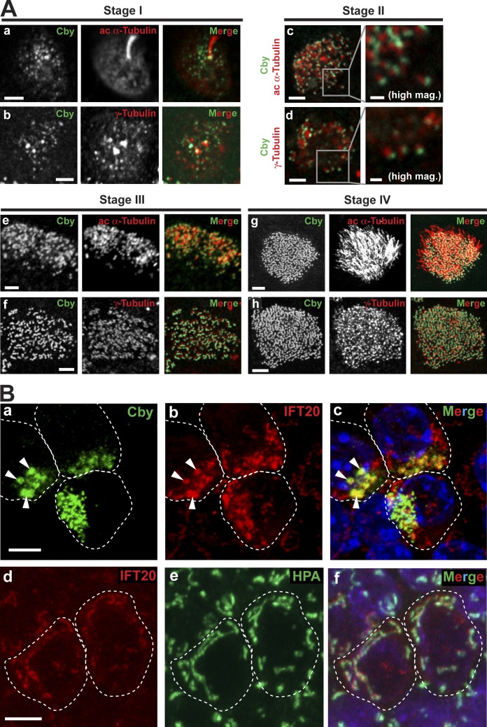
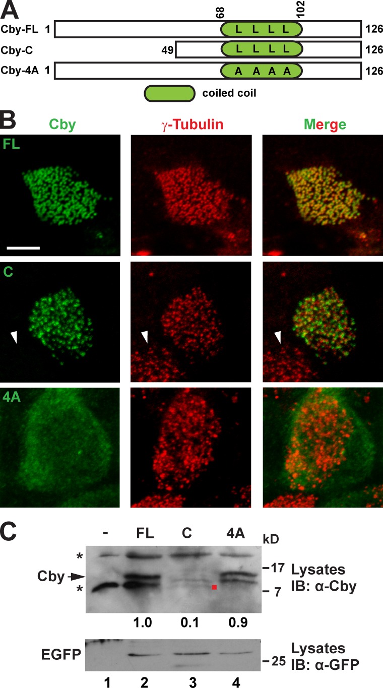
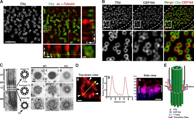
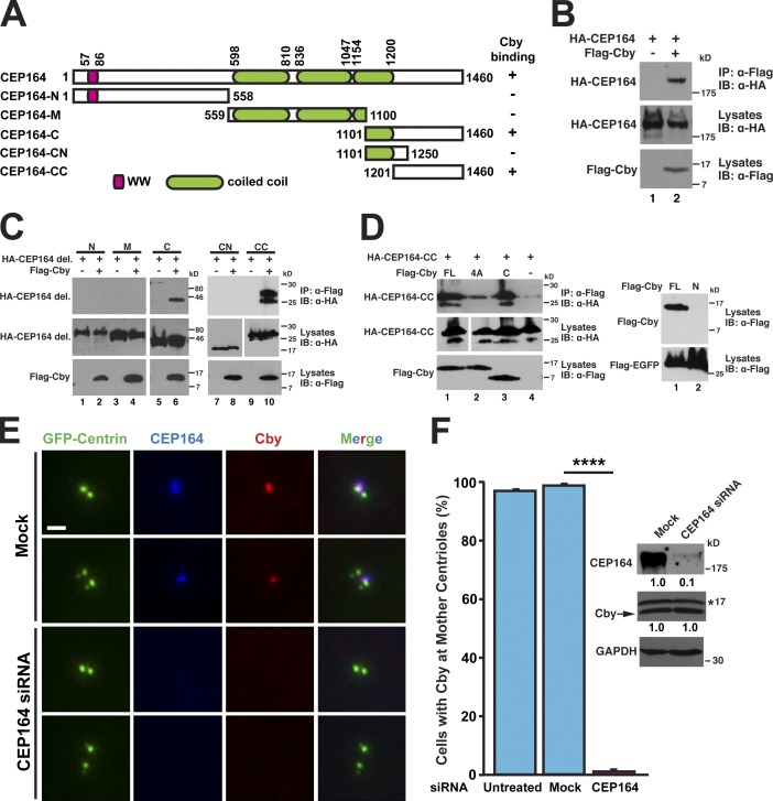
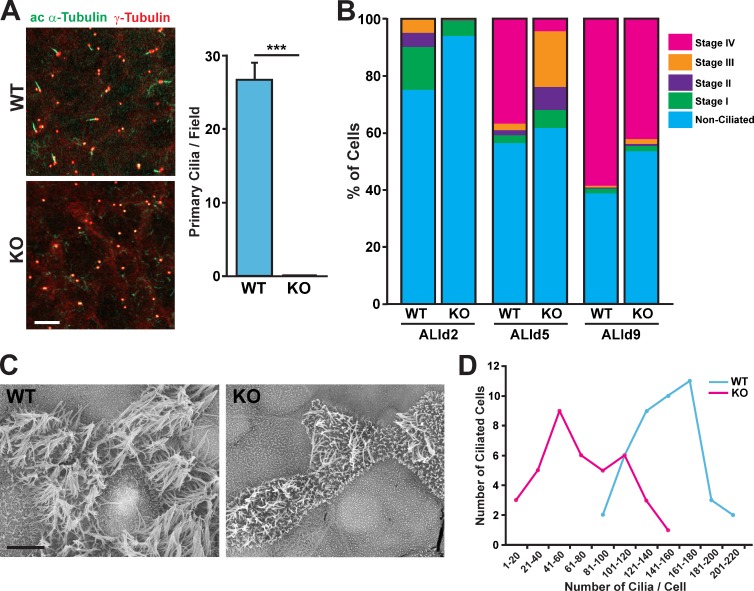
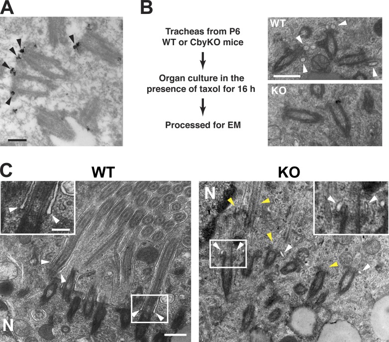
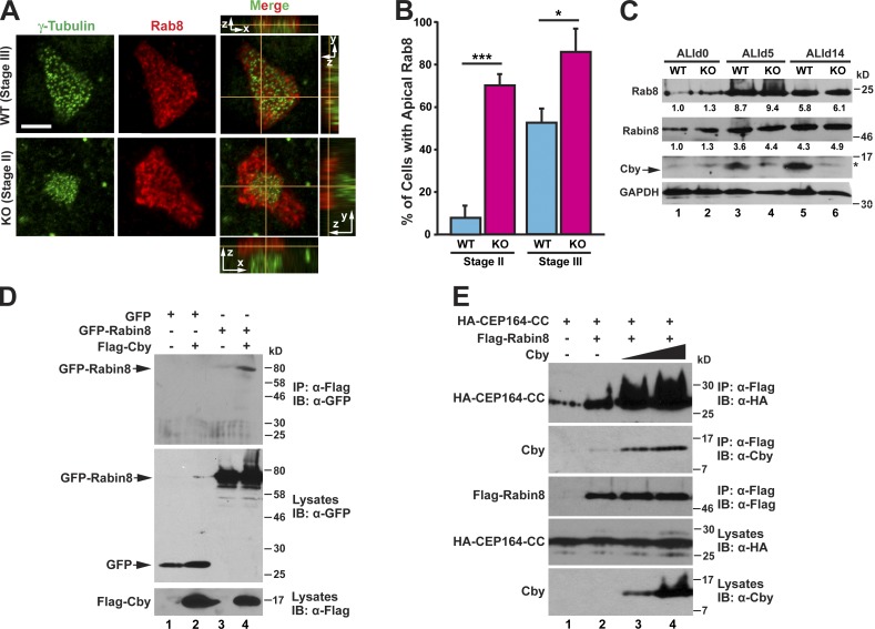
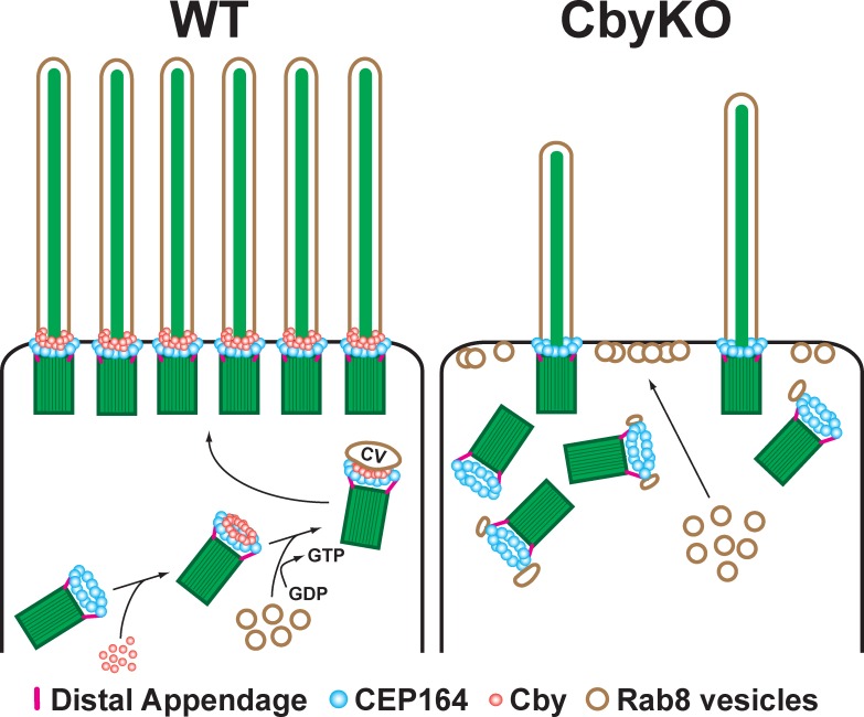
Airway multiciliated epithelial cells play crucial roles in the mucosal defense system, but their differentiation process remains poorly understood. Mice lacking the basal body component Chibby (Cby) exhibit impaired mucociliary transport caused by defective ciliogenesis, resulting in chronic airway infection. In this paper, using primary cultures of mouse tracheal epithelial cells, we show that Cby facilitates basal body docking to the apical cell membrane through proper formation of ciliary vesicles at the distal appendage during the early stages of ciliogenesis. Cby is recruited to the distal appendages of centrioles via physical interaction with the distal appendage protein CEP164. Cby then associates with the membrane trafficking machinery component Rabin8, a guanine nucleotide exchange factor for the small guanosine triphosphatase Rab8, to promote recruitment of Rab8 and efficient assembly of ciliary vesicles. Thus, our study identifies Cby as a key regulator of ciliary vesicle formation and basal body docking during the differentiation of airway ciliated cells.


Figures

Figure 1.
Figure 1.
Cby localization at different stages of ciliated cell differentiation. (A) MTECs were colabeled at the indicated ciliogenesis stages with antibodies against Cby and the centriole/basal body/ciliary axoneme marker acetylated (ac) α-tubulin or the centriole/basal body marker γ-tubulin. Essentially, immunostaining of CbyKO MTECs with the Cby antibody produced no signals (not depicted) confirming the antibody specificity. Bars: (main images) 2 µm; (high magnification images for stage II) 0.5 µm. See also Fig. S2 A. (B) Stage II MTECs were costained with Cby and IFT20 antibodies (a–c) or with the IFT20 antibody and the cis-medial Golgi marker lectin HPA (d–f). Nuclei in the merged image were visualized by DAPI (blue). (a–c) Arrowheads indicate areas of diffuse Cby staining that prominently overlap with IFT20. (d–f) In ciliated cells, IFT20 also showed Golgi localization. Dashed lines demarcate cell boundaries. Bars, 5 µm. See also Fig. S2 B.
Figure 2.
Figure 2.
The C-terminal coiled-coil motif is essential for Cby basal body localization. (A) Schematic depiction of human Cby constructs used in this study. The numbers indicate amino acid positions. Four leucines at positions 77, 84, 91, and 98 crucial for Cby homodimerization are shown. The dimerization-defective Cby-4A mutant contains alanine substitutions for all four leucine residues. (B) CbyKO MTECs were infected with lentiviruses encoding human full-length Cby (FL), Cby-C containing a leucine zipper coiled-coil motif, or Cby-4A mutant and colabeled with Cby and γ-tubulin antibodies at ALId14. Note that no Cby fluorescence was seen in noninfected ciliated cells (arrowheads). Bar, 5 µm. (C) Western blotting for expression of Cby mutants. Cell lysates were prepared from uninfected or infected CbyKO MTECs at ALId14 and probed with the Cby antibody. Note that Cby-C was detected at low levels (red dot). The asterisks indicate nonspecific bands present in uninfected cell lysates. The lentiviral constructs expressed EGFP from internal ribosome entry site, and similar transduction rates were verified by Western blotting with the GFP antibody. The band intensity of Cby proteins was quantified and normalized to that of EGFP. The normalized value for FL-Cby was set as 1. IB, immunoblot.
Figure 3.
Figure 3.
Cby protein is enriched at transition fibers and in the proximal compartment of the transition zone of mature cilia. (A) Fully differentiated MTECs at ALId14 were colabeled with antibodies for Cby and the ciliary axoneme marker acetylated (ac) α-tubulin and imaged by superresolution 3D-SIM. Top-down (x-y; single sections) and side (y-z on the right and x-z on the bottom; maximum projections) views are shown. The squared area is shown in higher magnification at the bottom right corner. See also Video 1. (B) Cby clusters as rings smaller than CEP164 rings at the ciliary base. Fully differentiated MTECs were costained with antibodies against Cby and CEP164 and imaged by 3D-SIM. The images are maximum projections from 10 z sections taken at 125-nm intervals. Top-down views and high-magnification views of the square areas are shown. Dashed lines demarcate cell boundaries. See also Fig. S3. (C) Immuno-EM localization of Cby in thin cross sections (80 nm) through the distal (b and c) and proximal (e and f) regions of the transition zone and the distal part of basal bodies (h and i) at the base of cilia in WT MTEC cultures at ALId14. (d, g, and j) CbyKO MTECs were processed in parallel as a negative control. A representative EM image of a cilium in longitudinal section is shown in a. Arrowheads in a denote the transition fiber. CP, cilia proper; BP, basal plate; TZ, transition zone; BB, basal body; TF, transition fiber. (D) Superresolution 3D-STORM images of Cby ring substructure in ALId14 MTEC cultures. Shown are a top-down view (a), intensity profile along the yellow arrow in a (b), and side view with a height map (c). White arrowheads in a point to Cby clusters. Brackets in c indicate the areas in which Cby molecules were highly enriched. The data are from a single representative experiment out of three repeats. (E) Cartoon depicting Cby localization at the base of mature cilia. Bars: (A, main image) 1 µm; (A [bottom right], C, and D) 0.1 µm.
Figure 4.
Figure 4.
CEP164 binds and recruits Cby to the distal appendages. (A) Schematic diagram of CEP164 constructs used in this study. The numbers indicate amino acid positions. (B) HEK293T cells were transfected with HA-CEP164 and Flag-Cby expression constructs as indicated, and cell lysates were immunoprecipitated with the Flag antibody and immunoblotted with the HA antibody. Cell lysates were also probed with HA and Flag antibodies to show stable protein expression. (C) Cell lysates from HEK293T cells expressing Flag-Cby and the indicated HA-tagged CEP164 fragments were immunoprecipitated with the Flag antibody and detected with the HA antibody. del., deletion. (D) CEP164 binds to the C-terminal region of Cby. HEK293T cells were transfected with expression constructs for HA–CEP164-CC and Flag-tagged Cby-FL, -4A, or -C (aa 64–126), and cell lysates were immunoprecipitated with the Flag antibody and immunoblotted with the HA antibody. (right) The N-terminal half of Cby (aa 1–63) was undetectable upon transfection. Cells were cotransfected with an expression plasmid for Flag-EGFP to normalize for transfection efficiency. White lines indicate that intervening lanes have been spliced out. (E) HeLa cells expressing GFP–centrin-1 were mock treated (all reagents except for siRNA) or transfected with CEP164 siRNA and immunostained with CEP164 and Cby antibodies. Cells with a pair or two pairs of centrioles are shown for each siRNA. Bar, 1 µm. (F) Quantification of the number of cells with Cby at the mother centrioles after siRNA treatment. U2OS cells were untreated, mock transfected, or transfected with CEP164 siRNA for 27 h and immunostained for Cby and CEP164. A total of 811 (untreated), 595 (mock), and 856 (CEP164 siRNA) cells were counted from at least five independent experiments. Data are presented as means ± SD. ****, P < 0.0001. Expression levels of CEP164, Cby, and GAPDH were determined by Western blotting. The asterisk indicates nonspecific bands. The band intensities of CEP164 and Cby were quantified and normalized to those of GAPDH. The normalized values for mock-transfected samples were set as 1. IB, immunoblot; IP, immunoprecipitation.
Figure 5.
Figure 5.
Impaired ciliogenesis in CbyKO ciliated cells. (A) WT and CbyKO MTECs were colabeled at ALId2 with acetylated (ac) α-tubulin and γ-tubulin antibodies. Bar, 10 µm. The quantification of primary cilia is shown in the graph on the right. For each genotype, primary cilia were counted in three microscopic fields (100× magnification) per MTEC preparation, and data are presented as means ± SEM from three independent MTEC preparations. ***, P < 0.001. (B) WT and CbyKO MTECs were fixed at ALId2, d5, and d9, and the percentage of cells in each stage of ciliogenesis were quantified based on immunostaining for acetylated α-tubulin and γ-tubulin. At least 600 cells/genotype/ALI day were counted from three independent MTEC preparations. See also Fig. S4. (C) Representative SEM images of WT and CbyKO ciliated cells in MTEC cultures at ALId9. Numerous short microvilli are apparent on the apical surface of CbyKO ciliated cells. Bar, 5 µm. (D) The number of cilia on ciliated cells was quantified on SEM images of MTEC cultures at ALId9, and the distribution of ciliated cells with numbers of cilia per cell was grouped into bins of 20. WT, n = 43; KO, n = 39.
Figure 6.
Figure 6.
Diminished recruitment of vesicles to the distal appendages in CbyKO ciliated cells. (A) Immuno-EM localization of Cby in early ciliated cell differentiation in MTEC cultures. Cby protein (arrowheads) localizes to the distal appendages of migrating basal bodies in the cytoplasm. See also Video 2. (B) Tracheal explants from P6 WT and CbyKO mice were cultured in the presence of taxol for 16 h to enrich vesicle-bound centrioles and processed for EM. Representative EM images are shown. Centrioles attached to vesicles (arrowheads) were more abundant in WT than CbyKO ciliated cells. (C) EM images of ciliated cells from taxol-treated P6 tracheas. The squared areas are shown in higher magnification at the top corners. In WT, cilium growth was occasionally observed in the cytoplasm, with ciliary membrane originating from the distal appendages (arrowheads). Although extended axonemal microtubules were present in CbyKO (yellow arrowheads), elongated ciliary membrane was not noticeable, but instead, centrioles were associated with small vesicles (white arrowheads). N, nucleus. Bars: (A and C, insets) 0.2 µm; (B and C, main images) 0.5 µm.
Figure 7.
Figure 7.
Cby facilitates recruitment of Rab8 to the basal bodies via stabilization of the CEP164–Rabin8 complex. (A) MTECs were fixed at ALId4 and immunostained for γ-tubulin and Rab8. Top-down (x-y) and side (y-z on the right and x-z on the top or bottom) views are shown. Bar, 5 µm. (B) Quantification of the number of ciliated cells with apical Rab8 at the indicated ciliogenesis stages. MTECs were fixed at ALId3 through d6 and colabeled with γ-tubulin and Rab8 antibodies. For each genotype, 51–108 ciliated cells were counted per MTEC preparation, and data are presented as means ± SEM from four independent MTEC preparations. *, P < 0.05; ***, P < 0.001. (C) Cell lysates were prepared from WT or CbyKO MTECs at ALId0, d5, and d14 and subjected to Western blotting for Rab8, Rabin8, Cby, and GAPDH as indicated. The asterisk indicates a nonspecific band. The band intensities of Rab8 and Rabin8 were quantified and normalized to those of GAPDH. The normalized values for WT samples at ALId0 were set as 1. (D) HEK293T cells were transfected with GFP or GFP-Rabin8 and Flag-Cby expression plasmids as shown, and cell lysates were immunoprecipitated with the Flag antibody and probed with the GFP antibody. (E) Cell lysates were prepared from HEK293T cells expressing HA–CEP164-CC, Flag-Rabin8, and increasing amounts of untagged Cby, which was immunoprecipitated with the Flag antibody and detected with the HA, Cby, or Flag antibody as shown. IB, immunoblot; IP, immunoprecipitation.
Figure 8.
Figure 8.
Model for basal body docking during airway ciliated cell differentiation. Upon centriole replication, CEP164 recruits Cby to the distal appendages. Cby in turn promotes efficient recruitment and fusion of Rab8-positive vesicles to form a ciliary vesicle at the distal end of centrioles. This facilitates docking of basal bodies to the apical membrane. In the absence of Cby, ciliary vesicles fail to form as a result of the inability to recruit sufficient Rab8 vesicles, resulting in compromised basal body docking. CV, ciliary vesicle. See Discussion for details.

Similar articles

Cited by

References

    1. Blatt, E.N., Yan X.H., Wuerffel M.K., Hamilos D.L., and Brody S.L.. 1999. Forkhead transcription factor HFH-4 expression is temporally related to ciliogenesis. Am. J. Respir. Cell Mol. Biol. 21:168–176 10.1165/ajrcmb.21.2.3691 - DOI - PubMed
    1. Boisvieux-Ulrich, E., Laine M.C., and Sandoz D.. 1989. In vitro effects of taxol on ciliogenesis in quail oviduct. J. Cell Sci. 92:9–20 - PubMed
    1. Chaki, M., Airik R., Ghosh A.K., Giles R.H., Chen R., Slaats G.G., Wang H., Hurd T.W., Zhou W., Cluckey A., et al. . 2012. Exome capture reveals ZNF423 and CEP164 mutations, linking renal ciliopathies to DNA damage response signaling. Cell. 150:533–548 10.1016/j.cell.2012.06.028 - DOI - PMC - PubMed
    1. Chang, J., Seo S.G., Lee K.H., Nagashima K., Bang J.K., Kim B.Y., Erikson R.L., Lee K.W., Lee H.J., Park J.E., and Lee K.S.. 2013. Essential role of Cenexin1, but not Odf2, in ciliogenesis. Cell Cycle. 12:655–662 10.4161/cc.23585 - DOI - PMC - PubMed
    1. Chiba, S., Amagai Y., Homma Y., Fukuda M., and Mizuno K.. 2013. NDR2-mediated Rabin8 phosphorylation is crucial for ciliogenesis by switching binding specificity from phosphatidylserine to Sec15. EMBO J. 32:874–885 10.1038/emboj.2013.32 - DOI - PMC - PubMed

Publication types

MeSH terms