. 2014 Oct 14;9(10):e110516.
doi: 10.1371/journal.pone.0110516. eCollection 2014.

Spontaneous proliferation of H2M-/- CD4 T cells results in unusual acute hepatocellular necrosis

Affiliations

Spontaneous proliferation of H2M-/- CD4 T cells results in unusual acute hepatocellular necrosis

Jeong-su Do et al. PLoS One. .

Abstract

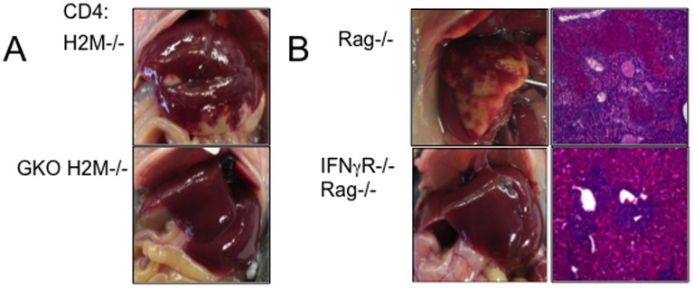
Naïve CD4 T cells are triggered to undergo spontaneous proliferation, a proliferative response induced in response to homeostatic stimulation, when exposed to severe lymphopenic environments. They spontaneously acquire proinflammatory effector phenotypes, playing a major role in inducing chronic inflammation in the intestine that is believed to be induced by T cell recognition of commensal antigens. While the antigens inducing the T cell responses and inflammation are being extensively investigated, the role of clonality of T cells involved in this process remains poorly understood. In this study, we utilized naïve CD4 T cells isolated from B6 H2M-/- mice, in which MHCII molecules are complexed with a single CLIP molecule, and examined spontaneous proliferation and intestinal inflammation of CD4 T cells expressing limited T cell receptor repertoire diversity. We found that H2M-/- CD4 T cells undergo robust spontaneous proliferation, differentiate into IFNγ-producing Th1 type effector cells, and, most unexpectedly, induce severe acute hepatocellular necrosis. T cell interaction with MHCII molecule on cells of hematopoietic origin was essential to induce the pathology. Interestingly, B cells are fully capable of preventing necrotic inflammation via IL-10-independent and B7-H1-dependent mechanism. This could be a useful animal model to examine T cell-mediated liver inflammation and B cell-mediated immune regulation.


Conflict of interest statement

Competing Interests: The authors have declared that no competing interests exist.

Figures

Figure 1
Figure 1. H2M−/− CD4 T cells induce severe necrotic inflammation in the liver.
WT and H2M−/− naïve CD4 T cells were adoptively transferred into Rag−/− recipients. (A) Weight loss and (B) survival were monitored after CD4 T cell transfer. Data are the mean ± SD of 4–9 individually tested animals from 2–3 independent experiments. (C) Liver picture and H&E stain of liver, (D) absolute number of donor CD4 T cells in different tissue, and (E) intracellular cytokine expression of the donor cells from the indicated tissues were determined 7 days post transfer. (F) Immunohistochemistry analysis of the CD3+ cells in the liver. Each symbol represents individually tested animals from two independent experiments. *, p<0.05; ***, p<0.001.
Figure 2
Figure 2. Distribution of TCR Vβ chain expression and liver inflammation induced by H2M−/− CD4 T cell subsets.
(A) TCR Vβ chain was measured at 7 days after transfer. Cells were isolated from SPL and stained with anti-TCR Vβ antibodies. Data are the mean ± SD of 5 individually tested animals from two independent experiments. (B) Subtype of TCR Vβ chain was measured from liver draining LN and mLN. Data are the mean ± SD of 4 individually tested animals. (C) Induction of liver inflammation by Vβ+ subtype H2M−/− CD4 T cells. FACS purified Vβ4, Vβ5/6, and Vβ8.1 H2M−/− CD4 T cells were transferred into Rag−/− mice and analyzed liver pathogenesis at day 7. (D) Absolute number of donor CD4 T cells in the spleen and liver. Data are the mean ± SD of 2–3 individually tested animals.
Figure 3
Figure 3. MHCII expression on recipients is required for liver inflammation mediated by H2M−/− CD4 T cells.
(A) Liver pathogenesis in Rag−/− and H2M−/− Rag−/− recipients after H2M−/− T cell transfer. (B) Absolute numbers of donor CD4 T cells were analyzed 7 days post transfer. (C) IFNγ and IL-17A cytokine expression were determined by FACS analysis. (D and E) Lethally irradiated MHCII−/− Rag−/− mice were reconstituted with BM cells from Rag−/− (Rag−/− BM → MHCII−/− Rag−/−, WT → KO). FACS purified naive H2M−/− CD4 T cells were transferred into the reconstituted recipients after 6 weeks of BM reconstitution. Recipients were sacrificed 7 days post transfer. (D) Liver pathology and (E) absolute numbers of donor cells were assessed. All experiments were repeated twice and similar results were observed. Each symbol represents individually tested animals. *, p<0.05; **, p<0.01.
Figure 4
Figure 4. B cells prevent liver necrotic inflammation.
(A) H2M−/− naïve CD4 T cells were transferred into Rag−/− and TCRβδ−/− recipients. Liver pathology and the absolute number of CD4 T cells in Rag−/− and TCRβδ−/− were examined at day 7. All experiments were repeated twice and similar results were observed. Each symbol represents individually tested animals. (B) Liver pathology and the absolute number of CD4 T cells in TCRβδ−/− and B cell depleted TCRβδ−/− recipients. Each symbol represents individually tested animals. *, p<0.05; **, p<0.01; ***, p<0.001.
Figure 5
Figure 5. MHCII and B7-H1 on B cells are required to prevent liver inflammation.
(A) H2M−/− naïve CD4 T cells were adoptively transferred into Rag−/− recipients together with 5×10⁶ WT, IL-10−/−, and B7-H1−/− B cells. Liver pathology and H&E stain (A) and the absolute numbers of CD4 T cells (B) in the liver were assessed 7 days after transfer. (C) H2M−/− naïve CD4 T cells were transferred into Rag−/− recipients. TCRβδ−/− serum was intravenously injected. (D) H2M−/− naïve CD4 T cells were adoptively transferred into Rag−/− recipients together with 5×10⁶ MHCII−/− B cells. All experiments were repeated twice and similar results were observed. Each symbol represents individually tested animals.
Figure 6
Figure 6. IFNγ is a key mediator of liver inflammation.
(A) H2M−/− and GKO H2M−/− naïve CD4 T cells were adoptively transferred into Rag−/− recipients. (B) H2M−/− naïve CD4 T cells were adoptively transferred into Rag−/− and IFNγR−/− Rag−/− recipients. Liver pathology was determined at 7 days post transfer. Representative pictures from three independent recipients are shown.

References

    1. Goldrath AW, Bevan MJ (1999) Selecting and maintaining a diverse T-cell repertoire. Nature 402: 255–262. - PubMed
    1. Do JS, Foucras G, Kamada N, Schenk AF, Shaw M, et al. (2012) Both exogenous commensal and endogenous self antigens stimulate T cell proliferation under lymphopenic conditions. Cell Immunol 272: 117–123. - PMC - PubMed
    1. Brimnes J, Reimann J, Nissen M, Claesson M (2001) Enteric bacterial antigens activate CD4(+) T cells from scid mice with inflammatory bowel disease. European journal of immunology 31: 23–31. - PubMed
    1. Powrie F, Leach MW, Mauze S, Menon S, Caddle LB, et al. (1994) Inhibition of Th1 responses prevents inflammatory bowel disease in scid mice reconstituted with CD45RBhi CD4+ T cells. Immunity 1: 553–562. - PubMed
    1. Viret C, Wong FS, Janeway CA Jr (1999) Designing and maintaining the mature TCR repertoire: the continuum of self-peptide:self-MHC complex recognition. Immunity 10: 559–568. - PubMed

Publication types

MeSH terms

LinkOut - more resources