

Selective use of sequential digital dermoscopy imaging allows a cost reduction in the melanoma detection process: a belgian study of patients with a single or a small number of atypical nevi
- PMID: 25313898
- PMCID: PMC4196852
- DOI: 10.1371/journal.pone.0109339
Selective use of sequential digital dermoscopy imaging allows a cost reduction in the melanoma detection process: a belgian study of patients with a single or a small number of atypical nevi
Abstract
Background: Dermoscopy is a technique which improves melanoma detection. Optical dermoscopy uses a handheld optical device to observe the skin lesions without recording the images. Sequential digital dermoscopy imaging (SDDI) allows storage of the pictures and their comparison over time. Few studies have compared optical dermoscopy and SDDI from an economic perspective.
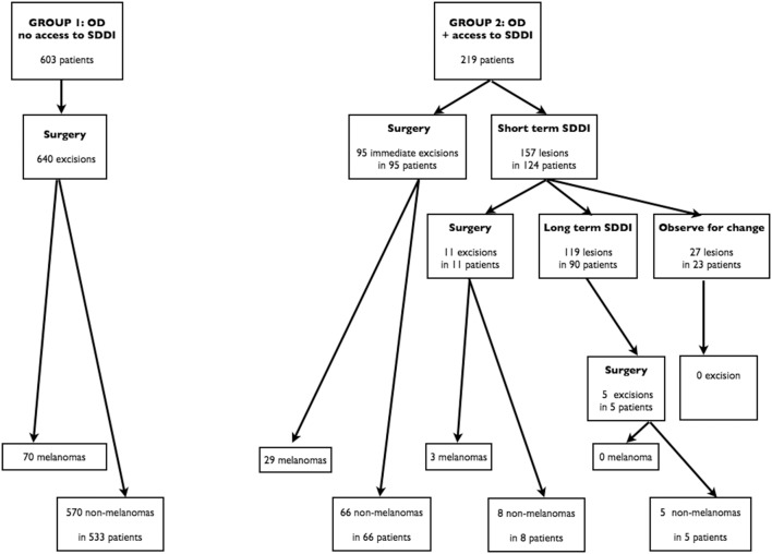
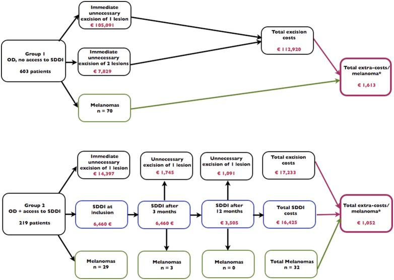
Objective: The present observational study focused on patients with one-to-three atypical melanocytic lesions, i.e. lesions considered as suspicious by optical dermoscopy. It aimed to calculate the "extra-costs" related to the process of melanoma detection. These extra-costs were defined as the costs of excision and pathology of benign lesions and/or the costs of follow-up by SDDI. The objective was to compare these extra-costs when using optical dermoscopy exclusively versus optical dermoscopy with selective use of SDDI.
Methods: In a first group of patients, dermatologists were adequately trained in optical dermoscopy but worked without access to SDDI. They excised all suspicious lesions to rule out melanoma. In a second group, the dermatologists were trained in optical and digital dermoscopy. They had the opportunity of choosing between immediate excision or follow-up by SDDI (with delayed excision if significant change was observed). The comparison of extra-costs in both groups was made possible by a decision tree model and by the division of the extra-costs by the number of melanomas diagnosed in each group. Belgian official tariffs and charges were used.
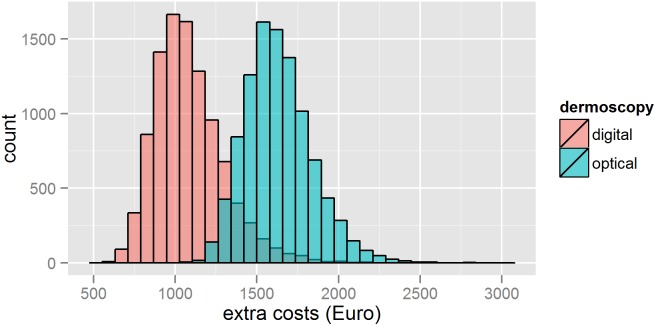
Results: The extra-costs in the first and in the second group were respectively €1,613 and €1,052 per melanoma excised. The difference was statistically significant.
Conclusions: Using the Belgian official tariffs and charges, we demonstrated that the selective use of SDDI for patients with one-to-three atypical melanocytic lesions resulted in a significant cost reduction.
Conflict of interest statement
Figures
References
-
- SEER Cancer statistics: Available: http://seer.cancer.gov/csr/1975_2010/Accessed 10 May 2013.
-
- Erdmann F, Lortet-Tieulent J, Schuz J, Zeeb H, Greinert R, et al. (2013) International trends in the incidence of malignant melanoma 1953–2008–are recent generations at higher or lower risk? Int J Cancer 132: 385–400. - PubMed
-
- Vestergaard ME, Macaskill P, Holt PE, Menzies SW (2008) Dermoscopy compared with naked eye examination for the diagnosis of primary melanoma: a meta-analysis of studies performed in a clinical setting. Br J Dermatol 159: 669–676. - PubMed
-
- Kittler H, Pehamberger H, Wolff K, Binder M (2002) Diagnostic accuracy of dermoscopy. The Lancet Oncology 3: 159–165. - PubMed
-
- Altamura D, Avramidis M, Menzies SW (2008) Assessment of the optimal interval for and sensitivity of short-term sequential digital dermoscopy monitoring for the diagnosis of melanoma. Arch Dermatol 144: 502–506. - PubMed
Publication types
MeSH terms
LinkOut - more resources
Full Text Sources
Other Literature Sources
Medical
