

Dental arch response to Haas-type rapid maxillary expansion anchored to deciduous vs permanent molars: A multicentric randomized controlled trial
- PMID: 25314034
- PMCID: PMC8611738
- DOI: 10.2319/041114-269.1
Dental arch response to Haas-type rapid maxillary expansion anchored to deciduous vs permanent molars: A multicentric randomized controlled trial
Abstract
Objective: To assess maxilla and mandibular arch widths' response to Haas-type rapid maxillary expansion (RME) anchored to deciduous vs permanent molars on children with unilateral posterior crossbite.
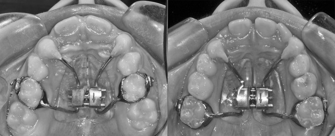
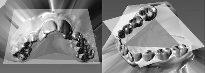
Materials and methods: Seventy patients with unilateral posterior crossbite recruited at the Universities of Genova, Siena, and Insubria (Varese) were randomly located into GrE (RME on second deciduous molars) or Gr6 (RME on first permanent molars) and compared.
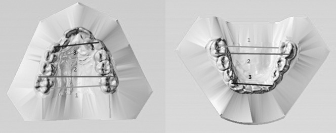
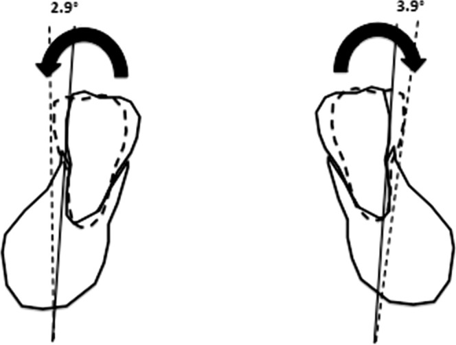
Results: Upper intermolar distance and permanent molar angulation increased significantly in Gr6 vs GrE at T1. Upper intercanine distance increased significantly in GrE vs Gr6 at T1 and T2. GrE showed significant increases for upper intermolar and upper intercanine widths. Gr6 showed statistically significant increases for upper intermolar widths, for upper and lower intercanine widths, and for increases of angulation of upper and lower permanent molars.
Conclusions: GrE showed reduced molar angulation increases at T1 and reduced molar angulation decreases at T2 when compared with Gr6. At T2, the net increase of the upper intercanine distance in GrE was still significant compared with Gr6, indicating a more stable expansion in the anterior area.
Keywords: Deciduous vs permanent molars; Multicentric randomized trial; Rapid maxillary expansion; Three-dimensional.
Figures
References
-
- McNamara J., Jr Maxillary transverse deficiency. Am J Orthod Dentofacial Orthop. 2000;117:567–570. - PubMed
-
- Thilander B, Wahlund S, Lennartsson B. The effect of early interceptive treatment in children with posterior cross-bite. Eur J Orthod. 1984;6:25–34. - PubMed
-
- Santos Pinto A, Buschang PH, Throckmorton GS, Chen P. Morphological and positional asymmetries of young children with functional unilateral posterior crossbite. Am J Orthod Dentofacial Orthop. 2001;120:513–520. - PubMed
-
- Petrén S, Bondemark L, Soderfeldt B. A systematic review concerning early orthodontic treatment of unilateral posterior crossbite. Angle Orthod. 2003;73:588–596. - PubMed
-
- Lagravere MO, Heo G, Major PW, Flores-Mir C. Meta-analysis of immediate changes with rapid maxillary expansion treatment. J Am Dental Assoc. 2006;13:44–53. - PubMed
Publication types
MeSH terms
LinkOut - more resources
Full Text Sources
Other Literature Sources
